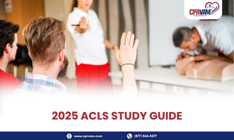
Many new healthcare providers consider obtaining ACLS certification, but they often back out due to its vast and complex course structure. One must have BLS certification to be eligible for ACLS training. The AHA continually updates its guidelines based on the latest scientific evidence, new research findings, and advancements in technology. The last one released was in 2020.
Here, we present you with a comprehensive ACLS study guide based on the latest AHA guidelines. This aims to support you in being ACLS certified. So, whether you are a first-time learner or getting the recertification after two years, this guide is going to be helpful for all learners. We have covered every key topic, like BLS review, ECG rhythm recognition, pharmacology, team dynamics, case scenarios, special considerations, and test practices in separate sections to help you understand with ease.
Let’s dive into the guide!
1. Advanced Cardiovascular Life Support (ACLS)
ACLS is the advanced life support administered to the victims of cardiovascular patients. Health professionals, who are involved in such critical and advanced care, require this skill to be able to treat the patient. ACLS for them is not just to meet their work standard, but it is crucial to be a responsible health professional and a caring individual as well.
ACLS adds to the BLS skills, ensuring a strong foundation in emergency handling and, at the same time, making health professionals proficient in complex cardiovascular emergencies. It includes key treatments like cardiac arrest, stroke, and acute coronary syndromes (ACS). It equips health professionals with in-depth knowledge of advanced cardiovascular conditions with the help of algorithms containing a step-by-step guide for treating patients.
2. Review of Basic Life Support (BLS) Skills
Basic Life Support (BLS) is the foundation of life-saving skills, which is the immediate first step in any real emergency. The chain of survival begins with administering basic life support. Here are the key components under BLS:
- First Aid Treatment: Provide immediate care to control bleeding, prevent shock, and maintain the airway before advanced help arrives.
- Chest Compressions: Perform hard and fast compressions at a depth of 2 inches and a rate of 100-120 per minute to maintain adequate blood circulation.
- Rescue Breaths: Deliver two effective rescue breaths after every 30 compressions, such that the chest rises with every breath.
- Early Recognition of Warnings: Identify signs of cardiac arrest or respiratory failure quickly to start BLS without delay.
- Automated External Defibrillator (AED) Usage: Use the AED promptly to analyze heart rhythm and deliver shocks if indicated to restart the heart or to restore normal rhythm.
- Teamwork: Coordinate roles efficiently among rescuers to minimize interruptions and optimize resuscitation.
- Post-ROSC Care: Provide appropriate medical intervention, supportive care, and constant monitoring after return of spontaneous circulation to prevent recurrence.
3. Initial Intervention
Resuscitation attempt should always begin with the assessment of the victim’s condition. The key technique is the ABC approach, which is checking airway, breathing, and circulation. The initial intervention is to ensure a patent airway, adequate ventilation, and effective circulation to maintain oxygen delivery to vital organs and prevent further damage.
ACLS trains the healthcare providers for the use of basic airway management tools like oropharyngeal (OPA) and nasopharyngeal (NPA), and advanced tools like endotracheal tubes (ETT), laryngeal mask airways (LMA), and i-gel devices. It also provides hands-on training for providing IV/IO access for medication delivery.
The next step should be to identify and treat the underlying and reversible causes of the cardiac arrest. Evaluate clinical signs, monitor data, and diagnostic tools to differentiate between potential causes of the patient’s collapse or deterioration. This process involves systematically considering the reversible causes (Hs and Ts) mnemonic for the identification of reversible causes. They are:
- Hs: Hypoxia, Hypovolemia, Hydrogen ion (acidosis), Hypo or hyperkalemia, Hypothermia
- Ts: Tension pneumothorax, Tamponade, Toxins, Thrombosis (pulmonary or coronary), and Trauma.
4. Pharmacology in ACLS
After recognizing the signs and symptoms of cardiac emergencies, initial ABC stabilization, and identifying the underlying and reversible causes, appropriate and timely medication therapy is essential. The pharmacology in ACLS includes the following drugs:
1. Epinephrine
- Used in cardiac arrest (e.g., ventricular fibrillation, pulseless ventricular tachycardia, asystole, PEA)
- Dose: 1 mg IV/IO every 3-5 minutes during resuscitation
2. Amiodarone
- Used for ventricular fibrillation or pulseless ventricular tachycardia resistant to shock and epinephrine
- Initial dose: 300 mg IV/IO bolus
- Repeat dose: 150 mg IV/IO if needed
3. Atropine
- Used for symptomatic bradycardia (slow heart rate)
- Dose: 0.5 mg IV every 3-5 minutes, max dose 3 mg
4. Adenosine
- Used for supraventricular tachycardia (SVT)
- Dose: 6 mg rapid IV push, if no conversion, then 12 mg IV push after 1-2 minutes
5. Magnesium Sulfate
- Used for torsades de pointes and hypomagnesemia
- Dose: 1-2 g IV diluted in 10 mL D5W over 5-20 minutes
6. Dopamine
- Used for bradycardia and hypotension when atropine is ineffective
- Dose: 2-20 mcg/kg/min IV infusion, titrated to response
7. Norepinephrine
- Used for severe hypotension and shock, including septic shock
- Dose: 0.1-0.5 mcg/kg/min IV infusion
8. Oxygen
Administer supplemental oxygen to maintain oxygen saturation of 90-99%
9. Aspirin
- For the acute coronary syndrome, to reduce clot formation
- Dose: 160-325 mg orally, chew if possible
10. Nitroglycerin
Used in acute coronary syndromes to reduce chest pain
5. ACLS Algorithms
There are systematic approaches to treat different cardiovascular conditions, given by individual algorithms for each of them. Health professionals gain hands-on practice for effective management and treatment based on these algorithms, compliant with the latest protocols and guidelines released by AHA.
Here are the algorithms in the ACLS course:
ACLS Cardiac Arrest Algorithm
ACLS includes the Cardiac arrest treatment algorithm, to treat the cardiovascular condition seen mainly in adults. The cardiac arrest is mainly categorized into two major rhythms:
- Shockable Rhythms (VF/pVT): The cardiac arrest characterized by shockable rhythms is treated starting with administering an electric shock, followed by continued CPR and constant checking of pulse or rhythm.
- Non-Shockable Rhythms (Asystole/PEA): In non-shockable rhythms, defibrillation is not given, but perform high-quality CPR immediately for 2 minutes straight without interruptions and keep checking the pulse. If still the same, proceed with the IV/IO access and then administer 1 mg epinephrine as soon as possible and repeat every 3–5 minutes. To support the ongoing intervention, identify and treat the reversible causes to prevent further complications.
ACLS Bradycardia Algorithm
The ACLS bradycardia algorithm helps healthcare professionals to treat symptomatic bradycardic patients. It includes:
- Recognition of early signs and symptoms, such as hypotension, pale skin, altered mental condition, chest pain, or acute heart failure in severe cases.
- Administer Atropine at a dose of 1 mg IV every 3–5 minutes, limiting up to 3 mg.
- If Atropine is ineffective, provide dopamine of 5 to 20 mcg/kg per minute or Epinephrine of 2 to 10 mcg per minute.
- Identification of underlying causes to support early and appropriate intervention.
ACLS Tachycardia Algorithm
Tachycardia treatment is based on whether the patient is stable or not. Specialized medical intervention is administered after assessment of stability. Here is how the algorithm works:
Unstable Tachycardia
- Administer synchronized cardioversion immediately.
Stable Tachycardia
- Check the QRS complex width and rhythm.
- For narrow QRS, administer vagal maneuvers and if not effective, provide 6 mg rapid IV push and 12 mg if needed.
- For a wide QRS, administer antiarrhythmic drugs like procainamide (20-50 mg/min IV), amiodarone (150 mg IV over 10 minutes).
- Immediately address any underlying causes.
Acute Coronary Syndrome (ACS)
The Acute Coronary Syndrome (ACS) algorithm in ACLS course trains the health professionals to provide step-by-step treatment by early recognition of the symptoms, initial stabilization, ECG readings, and administering appropriate medical intervention.
• Indicated by the 12-lead ECG.
• For NSTEMI, administer MONA (Morphine, Oxygen, Nitroglycerin.
• For STEMI, initiate Percutaneous Coronary Intervention (PCI) or Fibrinolytic Therapy.
Stroke
The ACLS algorithm for the treatment of stroke involves:
- Rapid identification of symptoms by recognizing sudden changes,
- Comprehensive assessment of neurological condition with brain imaging, laboratory tests, and analysis,
- Administer thrombolytic therapy
- Continuous monitoring and support.
6. ECG Rhythm Recognition
Key ECG Components for Rhythm Recognition
- P Wave: Atrial depolarization.
- QRS Complex: Ventricular depolarization.
- T Wave: Ventricular repolarization.
- PR Interval: Time from atrial to ventricular activation; critical in heart block diagnosis.
- Wide QRS: ≥ 0.12 seconds
- Narrow QRS: < 0.12 seconds
- Regularity: Regular or irregular rhythm aids in diagnosis.
Heart Rhythms
- Sinus Rhythm: Regular rhythm with normal P waves, QRS complexes, and T waves; rate 60-100 bpm.
- Bradycardia: Heart rate less than 60 bpm.
- Tachycardia: Heart rate over 100 bpm; P waves present, Supraventricular Tachycardia (SVT) if narrow complex and Ventricular Tachycardia (VT) if wide QRS complex originating from ventricles.
- Atrial Fibrillation (AF): No distinct P waves, irregularly irregular ventricular response.
- Ventricular Fibrillation (VF): Disorganized, chaotic electrical activity, no effective contraction.
- Asystole: No electrical activity visible, flatline.
- Pulseless Electrical Activity (PEA): Organized rhythm without a pulse.
Heart Block Levels
- First-Degree AV Block: Prolonged PR interval > 0.20 seconds, all atrial impulses are conducted to the ventricles.
- Second-Degree AV Block: when some atrial impulses fail to reach the ventricles
- Mobitz Type I (Wenckebach): Progressive lengthening of the PR interval until a QRS complex is dropped.
- Mobitz Type II: Intermittent dropped QRS without progressive PR lengthening; risk of progressing to complete heart block.
Third-Degree AV Block (Complete Heart Block): No relationship between P waves and QRS complexes; atrial and ventricular rhythms are independent
7. Team Dynamics
- Defined Roles and Responsibilities: Assigning clear tasks to each member, like compressions, airway, medication administration, and monitoring.
- Effective Communication: Clear order with all-around acknowledgement and feedback with no chance of miscommunication.
- Effective Leadership: Direct with calmness, priority-focused, and maintaining the code of conduct.
- Knowledge Sharing: Provision to suggest critical information if it helps patient care.
- Situational Awareness: Constantly reassess patient status, rhythm, and interventions.
- Debriefing Post-Code: Review performance to identify strengths and areas for improvement in future treatments.
8. Case Scenarios
ACLS comprises the simulation study and practice of the following scenarios:
1. Cardiac Arrest with Ventricular Fibrillation (VF): A patient suddenly collapses, has no pulse, and is found to have VF on the monitor. What is the immediate action?
2. Symptomatic Bradycardia: Patient has a very slow heart rate, causing low blood pressure, confusion, or chest pain. What’s the first-line treatment for this case?
3. Tachycardia with Pulse: What are the medical interventions to treat a patient with a rapid heart rate and symptoms like chest pain or hypotension?
4. Stroke Recognition and Response: How to carry out the early recognition and treatment to reduce brain damage and improve outcomes?
9. Special Considerations
ACLS certification courses cover special considerations, which are crucial in any advanced cardiovascular emergencies to increase recovery outcomes. They are:
- Treatment of Reversible Causes: Identification and management of underlying causes should always be in priority.
- Post-Resuscitation Care: Focus on stabilizing hemodynamics, oxygenation, and neurological status. Use targeted temperature management if indicated.
- Patient-Group Specific Treatment: Adjust protocols for different groups of patients (such as pregnant women, pediatrics, adults) using specific algorithms.
- Team and Communication: Efficient team roles, closed-loop communication, real-time feedback, and mutual respect ensure the team dynamics essential for improved treatment outcomes.
10. Assessment Guide
The ACLS certification assessment is not that hard to crack if you follow the proper guidelines and format for the study. Here are a few tips for effective preparation for ACLS skills test:
- Practice with Case Scenarios: ACLS skills tests include advanced-level skills tests to prepare you for handling cardiovascular emergencies effectively. Learn all the different case scenarios with megacodes for preparedness.
- Follow Structured Format: Create and follow a structured study schedule to ensure consistent study. Use mnemonics to remember steps and formulas for ACLS algorithms.
- Take Practice Tests: Take multiple ACLS practice exams to familiarize yourself with the test format, including precourse self-assessments.
- Get Familiar with Protocols: Keep yourself updated with the latest ACLS guidelines released by AHA.
- Cover Study Resources: Check the syllabus, get and use all the available resources such as guides, course PDFs, posters, formulae charts, videos, and practice question papers.
- Focused and Healthy Study Routine: Study in a calm and learning environment at the same time, ensuring proper rest and nutrition.
- Teamwork and Discussion: Along with self-practice, take the tests as a team to practice team dynamics in a simulated environment.
Ease your ACLS Certification Journey!
This ACLS study guide aims to help you get your ACLS certification easily by preparing for training sessions and assessments. By now, you are familiar with ACLS, including its different algorithms like cardiac arrest, stroke, ACS, and medical interventions in real emergencies with simulated scenarios. With the right study format, updates on protocols, and consistent effort, you can be certified in ACLS with no worries.
If you are looking for a trusted training provider, trust CPR VAM and get enrolled in your nearby locations. We offer comprehensive AHA-compliant initial as well as refresher courses in-person, online, or blended formats and certification on the course completion day, valid up to 2 years. Our training follows the latest 2020–2025 AHA guidelines, ensuring you’re learning the most current protocols.
Get ACLS certified today and boost your confidence and competency in life-threatening emergencies.
References
1. Advanced Cardiovascular Life Support (ACLS) | American Heart Association | CPR.Heart.org
https://cpr.heart.org/en/cpr-courses-and-kits/healthcare-professional/acls
2. ACLS Precourse Self-Assessment | elearning | American Heart Association | CPR.Heart.org
https://elearning.heart.org/course/423
3. Advanced Cardiovascular Life Support (ACLS) Course Options. | American Heart Association | CPR.Heart.org
https://cpr.heart.org/en/courses/advanced-cardiovascular-life-support-course-options

