CPRvam Students
25,000+ students successfully trained annually

Adult Tachyarrhythmia With a Pulse Algorithm (2025 AHA update), formally known as the ACLS Tachycardia Algorithm, is a structured clinical approach for managing adults with rapid heart rhythms and a pulse. It helps assess hemodynamic stability, identify the arrhythmia type, and guide timely, appropriate emergency cardiovascular treatment decisions.

Tachycardia is generally defined as a resting heart rate greater than 100 beats per minute (bpm). In acute clinical scenarios, rates ≥150 bpm are more likely to indicate a clinically significant tachyarrhythmia that requires urgent evaluation and management. These abnormal rhythms can reduce cardiac output and may lead to hypotension, shock, chest pain, or altered mental status, especially when associated with hemodynamic instability.

Step-by-step treatment algorithms

1. Supraventricular Tachycardias (SVT)

  1. Vagal Maneuvers: Attempt vagal maneuvers like the Valsalva maneuver, carotid sinus massage, or cold stimulus to the face.
  2.  Adenosine: Administer adenosine as a rapid intravenous (IV) bolus. It’s often the first-line medication for SVT.
  3. Beta-Blockers or Calcium Channel Blockers: In stable cases, oral or IV beta-blockers (e.g., metoprolol) or calcium channel blockers (e.g., diltiazem) may be used.

2. Atrial Fibrillation (AF) and Atrial Flutter

  1. Rate Control: Use medications such as beta-blockers, calcium channel blockers, or digoxin to control increased heart rate.
  2. Rhythm Control: Administer anti-arrhythmic medications like amiodarone or flecainide to restore and maintain normal sinus rhythm.
  3. Anticoagulation: Consider anticoagulant therapy to prevent thromboembolic events in patients with Atrial fibrillation at risk.

3. Ventricular Tachycardia (VT)

  1. Stable VT: Administer anti-arrhythmic drugs like amiodarone or lidocaine.
  2. Unstable VT: Perform synchronized cardioversion using appropriate energy levels.
  3. Consider Magnesium: Consider IV magnesium sulfate in cases of torsades de pointes or suspected magnesium deficiency.

4. Sinus Tachycardia:

  1. Identify and Treat Underlying Cause: Address and manage the underlying condition causing sinus tachycardia, such as pain, fever, or anxiety.
  2. Beta-Blockers: Consider beta-blockers if appropriate for symptom control.
Adult tachycardia with pulse algorithm step-by-step guide

The AHA ACLS Tachycardia with a pulse algorithm gives a clear and step-by-step approach to assess and treat patients with a fast heart rate. Start with immediate basic support (airway, breathing, oxygen if hypoxemic), place the patient on a monitor, get IV access, and a 12-lead ECG as soon as possible.

Key Steps in the ACLS Tachycardia Algorithm:

Step 1: Initial Recognition and Assessment

Initial assessment determines whether tachycardia is clinically significant, distinguishes physiological from pathological causes, and identifies reversible triggers such as fever, pain, or dehydration before initiating ACLS-directed management.

  • Confirm heart rate >100 bpm (clinically significant often ≥150 bpm)
  • Differentiate sinus tachycardia from pathological tachyarrhythmia
  • Evaluate for reversible causes:
    1. Fever
    2. Pain or anxiety
    3. Hypovolemia/dehydration
    4. Physical exertion
  • Treat the underlying cause if the tachycardia is physiological

Step 2: Immediate Stabilization and Monitoring

Immediate stabilization prioritizes airway, breathing, and circulation while establishing monitoring, vascular access, ECG evaluation, and continuous observation to detect early deterioration and guide urgent management decisions.

  • Assess and maintain airway patency
  • Support breathing if required
  • Administer oxygen only if hypoxemic
  • Attach continuous cardiac monitoring
  • Establish IV access promptly
  • Obtain a 12-lead ECG as early as possible

Step 3: Identify Persistent Tachyarrhythmia Causing Instability

Hemodynamic assessment identifies signs of instability that indicate poor perfusion or cardiovascular compromise, guiding whether immediate electrical therapy or further diagnostic evaluation is required.

  • Check for signs of instability:
    1. Hypotension
    2. Altered mental status
    3. Signs of shock
    4. Ischemic chest discomfort
    5. Acute heart failure

If Unstable → Perform immediate synchronized cardioversion without delay to restore circulation.
If Stable → Continue with detailed rhythm evaluation and ECG-based classification.

Step 4: ECG Evaluation – QRS Width

ECG interpretation focuses on QRS duration to differentiate supraventricular from ventricular origins, which directly determines treatment strategy and urgency of intervention.

  • A narrow QRS (<0.12 sec) indicates supraventricular tachycardia originating above the ventricles.
  • Wide QRS (≥0.12 sec) suggests ventricular tachycardia or abnormal ventricular conduction pathways.
  • Narrow-complex tachycardia is managed using the supraventricular tachycardia (SVT) treatment pathway.
  • Wide-complex tachycardia is treated as ventricular tachycardia until proven otherwise.

Step 5: Management of Stable Narrow-Complex Tachycardia

Treatment aims to slow AV nodal conduction, restore sinus rhythm when possible, and maintain hemodynamic stability using vagal maneuvers and AV nodal–blocking medications.

  • Perform vagal maneuvers if the rhythm is regular
  • Administer adenosine as first-line pharmacologic therapy
  • Consider repeating the adenosine if the initial dose fails
  • Use beta-blockers or calcium channel blockers for rate control
  • Continuous ECG monitoring throughout treatment
  • Seek expert consultation if the rhythm persists or the diagnosis is uncertain

Step 6: Management of Unstable Tachycardia


Unstable tachycardia requires immediate synchronized cardioversion to restore perfusion and prevent collapse. Electrical shock is synchronized with the cardiac cycle and prioritized over medications in life-threatening situations.

  • Perform immediate synchronized cardioversion
  • Do not delay for diagnostic testing or medications
  • Provide sedation only if it does not delay shock
  • Repeat cardioversion if rhythm persists
  • Adenosine may be considered only in selected stable SVT cases

Step 7: Post-Conversion or Refractory Tachycardia Management

Post-treatment care focuses on identifying underlying causes, preventing recurrence, and escalating therapy if rhythm disturbances persist despite initial interventions.

  • Reassess vital signs and cardiac rhythm
  • Identify and correct reversible causes:
    1. Hypoxia
    2. Electrolyte imbalance
    3. Myocardial ischemia
  • Repeat synchronized cardioversion if needed
  • Initiate antiarrhythmic infusion if indicated
  • Maintain continuous monitoring
  • Consult cardiology for refractory cases

Step 8: Stable Wide-Complex Tachycardia Management

Stable wide-complex tachycardia is treated cautiously as ventricular tachycardia until proven otherwise, requiring antiarrhythmic therapy and close monitoring to prevent deterioration.

  • Treat as ventricular tachycardia until excluded
  • Administer antiarrhythmic infusion: Amiodarone or procainamide
  • Adenosine may be used diagnostically in regular rhythms
  • Avoid AV nodal blockers if the diagnosis is uncertain
  • Continuous ECG and blood pressure monitoring required
  • Early specialist consultation recommended

Antiarrhythmic drugs restore or control the rhythm in stable tachycardia. They must be carefully dosed and monitored to avoid hypotension, proarrhythmia, or conduction abnormalities during infusion therapy.
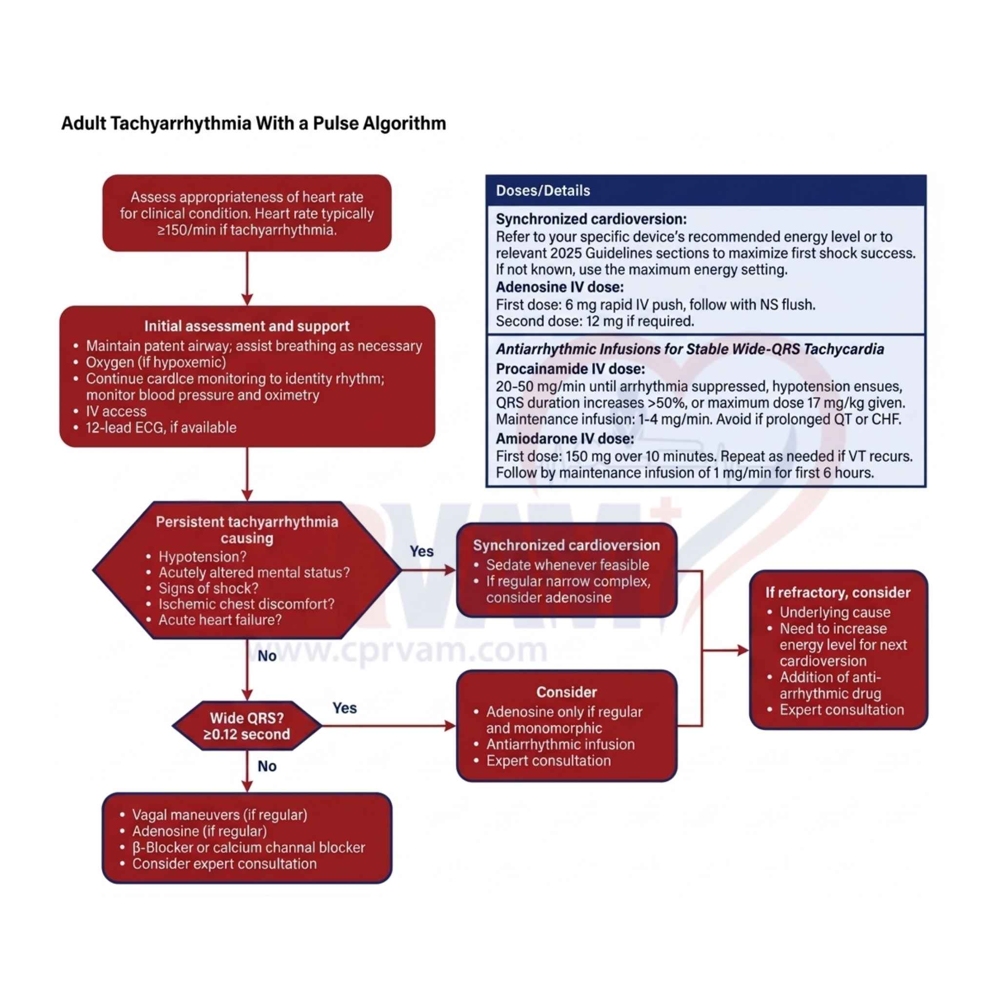
1. Adenosine

Adenosine briefly blocks AV node conduction to terminate regular narrow-complex SVT. It acts rapidly and has a very short duration, given as a fast IV bolus.

Essential Points:

  • Used for regular narrow-complex supraventricular tachycardia (SVT)
  • Given as 6 mg rapid IV push, then 12 mg if needed
  • Temporarily blocks AV node conduction to stop re-entry circuits
  • Causes brief flushing, chest discomfort, or transient pause

2. Amiodarone

Amiodarone is a broad-spectrum antiarrhythmic that stabilizes cardiac membranes and slows conduction. It is used in ventricular tachycardia and refractory supraventricular arrhythmias under monitoring.

Core Points:

  • Used for ventricular tachycardia and refractory SVT
  • Given as 150 mg IV over 10 minutes initially
  • Followed by maintenance infusion at 1 mg/min
  • May cause hypotension and bradycardia during infusion

3. Procainamide

Procainamide slows sodium channel conduction and prolongs refractoriness. It is mainly used for stable wide-complex tachycardia and requires slow infusion with careful monitoring.

Key Takeaways:

  • Used for stable wide-complex tachycardia (suspected VT)
  • Infused slowly at 20–50 mg per minute IV
  • Maximum total dose is 17 mg/kg
  • Avoid in prolonged QT interval and heart failure

4. Sotalol

Sotalol is a beta-blocker with class III antiarrhythmic effects. It prolongs repolarization and is used for stable ventricular and supraventricular tachycardias with ECG monitoring.

Important Points:

  • Used for stable ventricular and supraventricular tachycardias
  • Has both beta-blocking and potassium channel-blocking effects
  • Requires QT interval monitoring to prevent torsades de pointes
  • Administered in monitored clinical settings only

5. Beta-blockers (e.g., Metoprolol)

Beta-blockers reduce heart rate by blocking sympathetic stimulation and slowing AV node conduction. They are commonly used for rate control in stable tachycardias.

Main Points:

  • Used for rate control in stable tachycardia and atrial fibrillation
  • Example includes metoprolol given IV or orally
  • Works by blocking beta-adrenergic sympathetic activity
  • May cause bradycardia and hypotension if excessive

6. Calcium Channel Blockers (e.g., Diltiazem)

Diltiazem slows AV node conduction and reduces heart rate. It is used in stable supraventricular tachycardia and atrial fibrillation when beta-blockers are not suitable.

Key Points

  • Used for stable SVT and atrial fibrillation rate control
  • Works by blocking calcium channels in the AV node
  • Alternatively, when beta-blockers are contraindicated
  • May cause hypotension and bradycardia during use

The ACLS adult tachycardia algorithm offers a clear, step-by-step approach to assess and manage patients experiencing rapid heart rhythms. It is crucial to understand when to provide basic support, how to evaluate patient stability, and when to apply medications or electrical therapy to improve outcomes during tachycardia emergencies. Mastery of this algorithm ensures timely and effective treatment that can save lives.

The ACLS Tachycardia Algorithm is an essential tool for healthcare professionals aiming to build confidence and hands-on skills in managing tachycardia emergencies. By joining CPR VAM, you gain access to expert-led courses offering practical training and comprehensive knowledge, helping you become proficient in advanced cardiac life support. Start your learning journey today and be prepared to respond swiftly and effectively when every second counts.

The first treatment for unstable tachycardia is synchronized cardioversion. This is an electrical shock given at the right moment in the heartbeat to quickly restore a normal heart rhythm and improve symptoms. It is used when the fast heart rate is causing serious problems like low blood pressure or chest pain.

A heart rate over 150 beats per minute, especially with symptoms like chest pain, low blood pressure, or shortness of breath, may be dangerous and needs quick treatment.

Treatments may include giving oxygen, using medications to slow the heart rate, or applying electrical therapy like synchronized cardioversion if the patient is unstable.

It is used when an adult patient’s heart rate is faster than normal and is causing symptoms or could lead to serious problems.

Advanced Cadiovascular Life Support