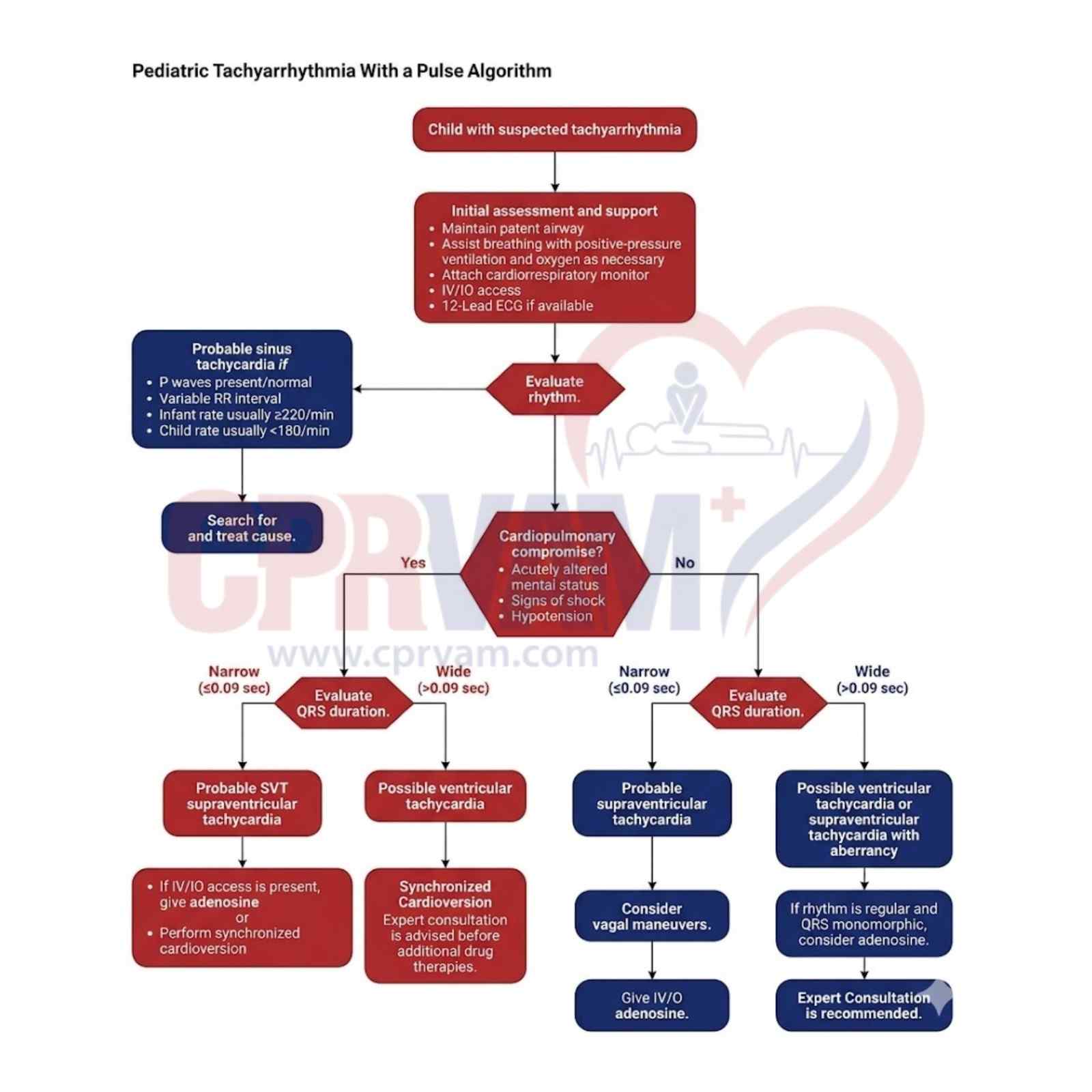
CPRvam Students
25,000+ students successfully trained annually
Step-by-step treatment algorithms

Effective management of pediatric tachycardia, guided by the American Heart Association 2025 approach, focuses on rapid rhythm identification, clinical assessment, and timely intervention. Providers must differentiate between stable and symptomatic cases, recognize underlying causes, and apply appropriate treatments to ensure safe, accurate, and outcome-driven pediatric emergency care.

Key Points:

  • Identify the specific tachycardia rhythm (e.g., sinus, SVT, atrial fibrillation)
  • Distinguish between narrow and wide complex tachycardia
  • Assess if the child has symptomatic tachycardia with a pulse
  • Evaluate overall clinical stability and perfusion status
  • Treat underlying or reversible causes promptly
  • Apply appropriate interventions, including cardioversion if indicated
Pediatric tachyarrhythmia with pulse algorithm flowchart

This structured AHA algorithm guides clinicians in assessing and treating kids with rapid heart rhythms who maintain a pulse. Emphasizes quick stabilization, rhythm analysis by QRS width, and interventions like adenosine or shocks to avert collapse.

This step starts when a child shows a very fast heart rate on symptoms or monitor readings, prompting immediate clinical assessment to quickly evaluate the condition and begin appropriate care.

Key Features:

  • Pallor, irritability, or visible distress
  • Rapid heart rate on monitor or palpation
  • Suspected abnormal tachycardia
  • Immediate activation of clinical response

Initial evaluation and support involve assessing the situation or condition at the beginning to identify key issues. It also includes providing immediate assistance to ensure safety, stability, and appropriate next steps.

Core Actions:

  • Ensure airway patency and adequate breathing
  • Provide oxygen if respiratory distress is present
  • Attach ECG and pulse oximetry monitors
  • Establish IV or IO access
  • Obtain a 12-lead ECG when available

Rhythm Analysis involves carefully reviewing the ECG along with the patient’s clinical signs. This helps differentiate normal sinus tachycardia from potentially dangerous tachyarrhythmias.

Assessment Focus:

  • Review the ECG/monitor rhythm carefully
  • Differentiate sinus tachycardia from arrhythmia
  • Assess perfusion and hemodynamic stability
  • Direct the patient to the appropriate treatment pathway

Probable sinus tachycardia is a normal physiological response to stress, fever, or volume loss, showing normal P waves and variable RR intervals.

Key Features:

  • Visible P waves
  • Rate appropriate for age:
    • Ā Infants: Usually <220 bpm
    • Children: Usually <180 bpm
  • Gradual onset and variability

3.1.1. Identify and Treat Underlying Cause

First, identify the underlying cause of tachycardia and provide appropriate treatment by addressing the condition responsible for helping restore normal heart rate and stability.

Clinical Actions:

  • Evaluate for fever, dehydration, pain, or infection
  • Treat the underlying cause promptly
  • Provide fluids or antipyretics as needed
  • Reassess heart rate after treatment

This step focuses on identifying whether the child’s condition is unstable and requires immediate action. Careful clinical evaluation helps detect early signs of serious deterioration.

Warning Signs:

  • Altered mental status
  • Signs of shock (poor perfusion, weak pulses)
  • Hypotension for age
  • Clinical deterioration

When warning signs are present, QRS duration is measured to help classify the tachyarrhythmia. This assessment guides diagnosis and supports timely, appropriate clinical management decisions.

Key Measurements:

  • Narrow QRS (≤0.09 sec): Measure QRS duration on ECG; if it is ≤0.09 seconds, it suggests a supraventricular origin of the tachyarrhythmia.
  • Wide QRS (>0.09 sec): If QRS duration is >0.09 seconds on ECG, it suggests a ventricular origin and requires closer evaluation.

Suspected supraventricular tachycardia (SVT) is a fast, regular heart rhythm originating above the ventricles, typically involving abnormal electrical activity in the atria or AV node.

Characteristic Features:

  • Sudden onset and termination
  • Regular rhythm
  • Absent or abnormal P waves
  • Narrow QRS complex

5.1.1. Management of SVT

Management of SVT involves a structured treatment approach aimed at restoring normal heart rhythm and stabilizing the patient.

Treatment Approach:

  • Administer adenosine via IV/IO access
  • If unstable or no access: perform synchronized cardioversion
  • Continuous cardiac monitoring required
  • Rapid intervention is essential

When an ECG shows a wide-complex tachycardia originating from the ventricles, it is identified as possible ventricular tachycardia (VT) and requires urgent assessment due to the high risk of deterioration.

Risk Features:

  • Wide-complex tachycardia
  • Ventricular origin suspected
  • Hemodynamic instability possible
  • Requires immediate management

5.2.1. Synchronized Cardioversion

Synchronized cardioversion is used to restore normal rhythm in unstable tachycardia, and expert consultation is recommended before initiating any additional drug therapy.

Shock Protocol:

  • Initial dose: 0.5-1 J/kg
  • Increase to 2 J/kg if ineffective
  • Sedate if time permits, and the patient is stable
  • Do not delay in unstable patients

In stable children without warning signs, QRS duration is evaluated to guide management decisions. Measuring QRS width helps determine the appropriate diagnostic and treatment pathway.

Reassessment:

  • Narrow QRS → SVT pathway
  • Wide QRS → VT pathway
  • Confirms correct treatment route

Confirmed supraventricular tachycardia is a stable, regular, fast heart rhythm with narrow QRS complexes seen on ECG. It usually starts above the ventricles and is managed with a specific treatment approach.

Features:

  • Narrow-complex tachycardia on ECG
  • Hemodynamically stable patient
  • Clinical and ECG findings consistent with SVT diagnosis

6.1.1. Vagal Maneuvers

Vagal maneuvers are the first-line, non-drug treatment for stable supraventricular tachycardia. They involve simple techniques that stimulate the vagus nerve to slow AV node conduction and may help terminate the abnormal rhythm and restore normal heart rate.

6.1.2. IV/IO Adenosine Administration

IV/IO adenosine administration is used for stable SVT, given rapidly through intravenous or intraosseous access to temporarily block AV node conduction and restore normal rhythm.

Dosing:

  • 0.1 mg/kg rapid IV/IO bolus (max 6 mg)
  • If ineffective: 0.2 mg/kg (max 12 mg)
  • Follow with a rapid saline flush

A wide-complex tachycardia may be due to either ventricular tachycardia or supraventricular tachycardia with aberrant conduction. Careful ECG review and clinical assessment are needed to correctly identify the rhythm and guide appropriate treatment.

6.2.1. Adenosine Trial

An adenosine trial is used in selected stable cases of wide-complex tachycardia as a diagnostic step to help distinguish supraventricular from ventricular origin.

Purpose:

  • Regular monomorphic wide-complex tachycardia
  • Helps differentiate SVT from VT
  • Must be done under monitoring

6.2.2. Specialist Consultation

Specialist consultation with pediatric cardiology is indicated when the heart rhythm is complex or uncertain. This step ensures expert evaluation, especially in difficult cases where diagnosis and management are not straightforward.

Support actions include consulting pediatric cardiology for guidance in complex or unclear rhythm cases. This collaboration improves diagnostic accuracy and enhances patient safety by ensuring appropriate interpretation and management decisions.

The PALS Tachycardia Algorithm (Pediatric Tachyarrhythmia with a Pulse Algorithm) is a structured, evidence-based clinical approach for rapidly assessing and managing children with fast heart rhythms who maintain a pulse. It guides healthcare providers through ABC evaluation, rhythm identification, stabilization, and timely interventions such as adenosine and synchronized cardioversion to prevent deterioration and improve outcomes.

This algorithm emphasizes early recognition, correction of reversible causes, continuous monitoring, and quick decision-making based on rhythm and stability. Healthcare professionals are encouraged to build competency through PALS training, which develops essential skills for pediatric emergencies. Structured programs like CPR VAM Training Center offer comprehensive certification courses, including BLS, PALS, and ACLS, for confident clinical practice.

In many cases, it can be triggered by common issues like fever, dehydration, pain, or emotional stress. More serious causes may include heart rhythm disorders or underlying heart conditions that need medical attention.

Doctors quickly check the child’s overall condition, including blood pressure, alertness, and circulation. If signs show instability or poor perfusion, it is treated as an emergency requiring immediate action.

The rhythm pattern helps determine whether the issue is coming from the upper or lower chambers of the heart. This distinction directly guides whether medications or electrical treatment may be needed.

Medical teams first focus on airway support, oxygen if needed, heart monitoring, and IV or IO access. At the same time, they try to identify and correct any reversible cause behind the rapid heart rate.

Stronger interventions are used when the child is unstable or when the rhythm does not respond to initial steps. These treatments are carefully controlled and aimed at quickly restoring a normal heart rhythm.

Pediatric Advanced Life Support (PALS) Classes