- 2026
CPR VAM • All Rights Reserved • Developed by Splendour Group

Understanding how to make critical decisions when every second matters
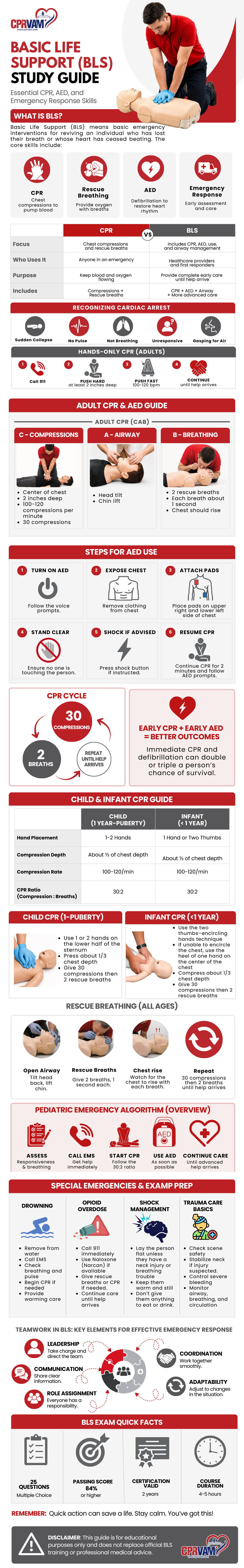
ACLS is more than following the algorithms. This guide helps you understand how to assess, prioritize, and respond during cardiovascular emergencies. The infographic provides a quick visual overview, while this resource supports ACLS review and serves as an ACLS study guide PDF for practical learning and revision.
Advanced Cardiovascular Life Support (ACLS) is an advanced emergency care approach used to assess, stabilize, and treat patients experiencing life-threatening cardiovascular emergencies.
Built on the foundation of Basic Life Support (BLS), ACLS combines advanced clinical interventions and structured decision-making to improve patient outcomes during critical situations.
ACLS Includes:
ACLS is commonly used in emergency departments, intensive care units, ambulances, and other acute care settings where rapid assessment and timely intervention are essential.
Every ACLS intervention begins with a strong foundation in Basic Life Support (BLS). Before advanced medications, airway procedures, or cardiac interventions are introduced, providers must first recognize emergencies, support circulation, and maintain oxygen delivery.
BLS focuses on immediate actions that help preserve life until advanced care can begin.
Strong BLS skills create the foundation for effective ACLS care and improve the success of advanced treatment.
High-quality CPR is one of the most important factors in improving survival during cardiac arrest. Effective chest compressions help maintain blood flow to the brain and other vital organs until normal heart function can be restored.
Delivering CPR correctly improves the effectiveness of advanced interventions and supports better patient outcomes.
Component | Recommended Target |
Compression Rate | 100–120 compressions per minute |
Compression Depth | 5–6 cm (2–2.4 inches) |
Chest Recoil | Allow complete recoil after each compression |
Compression Fraction | Greater than 80% with minimal interruptions |
Pulse Check | Keep under 10 seconds |
High-quality CPR creates the foundation for successful ACLS care and gives patients the best opportunity for recovery.
Before advanced treatment begins, every patient should be assessed using the ABC approach: Airway, Breathing, and Circulation. This structured method helps healthcare providers quickly identify the most immediate threats to life and deliver care in the correct order.
Airway refers to the passage that allows air to move into and out of the lungs. If the airway becomes blocked, oxygen cannot reach the body, making airway assessment the priority.
Providers assess whether the patient can move air effectively and whether additional airway support is needed.
During airway assessment, check for:
Common airway interventions:
Goal: Ensure oxygen can safely reach the lungs.
Once the airway is open, the next step is determining whether breathing is effective enough to deliver oxygen and remove carbon dioxide.
Breathing assessment focuses on both the quality and effectiveness of ventilation.
During breathing assessment, check for:
Common breathing interventions:
Goal: Maintain adequate oxygenation and effective ventilation.
Circulation determines whether oxygenated blood is reaching the brain, heart, and other vital organs.
Even if breathing is normal, poor circulation can rapidly become life-threatening.
During circulation assessment, check for:
Common circulation interventions:
Goal: Restore and maintain blood flow to support vital organ function.
The ABC Assessment creates the foundation for ACLS and guides every decision that follows during emergency care.
When a patient does not respond to resuscitation or remains clinically unstable, healthcare providers evaluate for reversible causes. In ACLS, these causes are grouped into Hs and Ts to help identify and treat underlying conditions that may be preventing recovery.
Recognizing and correcting these causes can improve the effectiveness of treatment and increase the likelihood of successful resuscitation.
These conditions affect the body’s normal function and may interfere with circulation, oxygen delivery, or heart activity.
Hypoxia
Low oxygen levels that reduce oxygen delivery to tissues and vital organs.
Hypovolemia
A significant loss of blood or body fluids that decreases circulating blood volume.
Hydrogen Ion (Acidosis)
An imbalance in the body’s acid levels can impair cardiac function.
Hypokalemia or Hyperkalemia
Low or high potassium levels can disrupt the normal electrical activity of the heart.
Hypothermia
A dangerously low body temperature that slows metabolism and affects cardiac performance.
These conditions are caused by physical obstruction, pressure changes, or harmful substances that interfere with normal heart and lung function.
Tension Pneumothorax
Air trapped in the chest places pressure on the lungs and reduces effective breathing.
Cardiac Tamponade
Fluid surrounding the heart restricts its ability to fill and pump properly.
Toxins
Medications, chemicals, or other substances that negatively affect cardiac or respiratory function.
Thrombosis (Pulmonary )
A blockage in the blood vessels of the lungs that limits oxygen exchange.
Thrombosis (Coronary)
A blockage in the coronary arteries that reduces blood flow to the heart muscle.
Identifying and treating reversible causes helps address the source of instability and supports better patient outcomes during ACLS care.
Medications play an important role in ACLS and are selected based on the patient’s heart rhythm, symptoms, and overall clinical condition. Understanding when and why each medication is used helps support faster and more effective decision-making during emergencies.
Medication | Common Use | Adult Dose |
Epinephrine | Used during cardiac arrest to improve blood flow during resuscitation | 1 mg IV/IO every 3-5 minutes |
Amiodarone | Used for shock-resistant ventricular fibrillation (VF) or pulseless ventricular tachycardia (VT) | 300 mg IV/IO |
Atropine | First-line medication for symptomatic bradycardia | 1 mg IV every 3-5 minutes |
Adenosine | Used to treat stable supraventricular tachycardia (SVT) | 6 mg rapid IV push, then 12 mg if needed |
Magnesium Sulfate | Used for Torsades de Pointes and magnesium-related rhythm disturbances | 1-2 g IV |
Dopamine | Supports heart rate and blood pressure when additional treatment is needed | Continuous IV infusion |
Norepinephrine | Used to manage severe hypotension and shock | Continuous IV infusion |
Aspirin | Used in Acute Coronary Syndrome (ACS) to reduce clot formation | 160-325 mg orally |
Nitroglycerin | Used to relieve chest pain associated with ACS | 0.4 mg sublingual every 5 minutes |
ACLS algorithms provide a structured, evidence-based approach to recognizing and managing cardiovascular emergencies. Rather than focusing only on memorizing treatment sequences, these algorithms guide healthcare professionals through assessment, intervention, reassessment, and clinical decision-making during time-critical situations.
Each algorithm is designed to support rapid action, improve team coordination, and deliver care based on the patient’s rhythm, symptoms, and overall condition.
Cardiac arrest occurs when the heart suddenly stops producing effective circulation, resulting in loss of pulse, reduced oxygen delivery, and immediate risk to life.
The first clinical decision is identifying whether the patient has a shockable or non-shockable rhythm, as this determines the treatment pathway.
Shockable rhythms are caused by abnormal electrical activity that may respond to defibrillation.
Recommended Management
Treatment Sequence
Non-shockable rhythms do not respond to defibrillation and require immediate circulation support and cause identification.
Recommended Management
Treatment Sequence
Bradycardia becomes clinically significant when a slow heart rate reduces perfusion and causes symptoms.
Treatment decisions should focus on whether the patient remains hemodynamically stable.
Assess for Signs of Instability
Recommended Management
Treatment Sequence
Tachycardia management depends on whether the patient remains clinically stable.
Assessment should focus on determining whether the rapid heart rate is compromising circulation.
Evaluate for Instability
If circulation remains stable:
If signs of poor perfusion are present:
Perform immediate synchronized cardioversion to restore stability.
Treatment Approach
Stable → Rhythm-based treatment
Unstable → Immediate cardioversion
Acute Coronary Syndrome includes conditions caused by reduced blood flow to the heart muscle and requires early recognition and timely intervention.
The primary goal is to restore perfusion and limit cardiac injury.
Initial Management
Goal
Restore blood flow quickly and reduce further myocardial damage.
Stroke is a medical emergency where early recognition and treatment directly influence outcomes.
Rapid assessment helps reduce neurological injury and improve recovery.
Recognition → Assessment → Imaging → Treatment → Monitoring
Goal
Recognize stroke early, confirm diagnosis quickly, and initiate timely treatment.
These ACLS algorithms support organized emergency care, clinical consistency, and informed decision-making during critical cardiovascular events.
ECG rhythm recognition is a fundamental ACLS skill that helps healthcare providers identify cardiac abnormalities quickly and make informed treatment decisions during emergencies.
An electrocardiogram (ECG) records the heart’s electrical activity and provides insight into heart rate, rhythm, and conduction. Understanding these patterns allows providers to recognize normal function, detect abnormalities early, and respond appropriately.
Before interpreting heart rhythms, it is important to understand the major ECG components and what they represent.
Represents Atrial Depolarization
The P wave reflects the electrical signal that causes the atria to contract and move blood into the ventricles.
When assessing the P wave, consider:
Abnormal or absent P waves may indicate atrial rhythm disturbances.
Represents Ventricular Depolarization
The QRS complex represents electrical activation of the ventricles and corresponds to ventricular contraction.
Evaluate:
QRS interpretation:
QRS width is an important factor when identifying arrhythmias and selecting treatment.
T Wave
Represents ventricular repolarization, the recovery phase after contraction.
Assess:
PR Interval
Represents the time required for electrical signals to travel from the atria to the ventricles.
Normal PR interval: 0.12–0.20 seconds
Changes in the PR interval may indicate delayed electrical conduction.
Rhythm regularity refers to whether heartbeats occur at consistent intervals.
Evaluate:
Regularity is often one of the first clues used to identify abnormal rhythms.
Normal sinus rhythm reflects normal electrical conduction through the heart.
Characteristics:
Clinical significance:
Indicates effective electrical activity and coordinated heart function.
Bradycardia is a slower-than-normal heart rate.
Characteristics:
Clinical Significance:
Treatment depends on symptoms and whether circulation is affected.
Tachycardia is a faster-than-normal heart rate.
Characteristics:
Clinical Significance:
Management depends on rhythm type and patient stability.
Atrial fibrillation occurs when atrial electrical activity becomes irregular and uncoordinated.
Characteristics:
Clinical significance:
Can reduce cardiac efficiency and requires clinical evaluation.
Ventricular fibrillation is a life-threatening rhythm that produces no effective cardiac output.
Characteristics:
Clinical significance:
Requires immediate defibrillation and high-quality CPR.
Asystole represents the absence of measurable cardiac electrical activity.
Characteristics:
Clinical significance:
Requires immediate resuscitation and evaluation for reversible causes.
Heart blocks occur when electrical signals between the atria and ventricles are delayed or interrupted.
Characteristics:
Clinical Significance:
Represents delayed conduction without missed ventricular beats.
Some atrial impulses fail to conduct to the ventricles.
Characteristics:
Characteristics:
Clinical Significance:
May progress to more advanced conduction disturbances.
Characteristics:
Clinical Significance:
Represents complete electrical dissociation and requires urgent clinical management.
ECG interpretation should always be combined with patient symptoms, clinical assessment, and ACLS protocols to support safe and effective decision-making.
Successful ACLS care depends on more than clinical knowledge and technical skills. Effective resuscitation requires healthcare teams to communicate clearly, work efficiently, and coordinate actions under pressure.
Strong team dynamics help reduce delays, improve patient safety, and support faster, more organized emergency care.
Exchange information clearly and efficiently
Communication during ACLS should be direct, concise, and focused on patient care. Team members should provide updates, communicate interventions, and report changes in patient status in real time.
Effective communication reduces delays, minimizes errors, and helps ensure the entire team remains aligned throughout resuscitation.
Assign clear roles to improve efficiency
Every team member should understand their responsibilities before and during resuscitation. Clearly assigned roles improve workflow and allow critical interventions to happen without duplication or confusion.
Common team roles may include:
Well defined responsibilities support a structured and efficient response.
Coordinate care and guide decision making
Strong leadership is essential during ACLS events. The team leader maintains oversight of the situation, prioritizes interventions, delegates tasks, and ensures treatment follows established protocols.
Effective leaders promote organization, maintain focus, and support timely clinical decision making.
Maintain awareness of the patient and the overall environment
Situational awareness means understanding both the patient’s condition and the actions taking place around the team.
Team members should continuously:
Maintaining awareness supports proactive decision making and more effective emergency care.
Encourage team input and clinical observations
Effective resuscitation relies on shared information. Every team member should communicate important findings, raise concerns, and provide updates throughout the event.
Knowledge sharing supports collaboration, strengthens decision making, and improves patient care.
Examples include:
Open communication helps teams respond more effectively.
Confirm instructions and completed actions
Closed loop communication ensures instructions are received, understood, and completed correctly.
The process includes:
This communication method improves accuracy, accountability, and team coordination.
Review performance and identify opportunities to improve
After resuscitation, the team should conduct a brief review to evaluate performance and identify areas for improvement.
Debriefing helps teams:
Continuous review supports long term improvement in emergency care delivery.
Effective ACLS combines clinical expertise with strong teamwork to support safe, efficient, and patient focused care.
Critical moments in an ACLS scenario that require timely assessment and informed clinical decisions. Use patient presentation, rhythm recognition, and ACLS algorithms to determine the most appropriate next step in patient care.
This is a life-threatening cardiac emergency where the heart has chaotic electrical activity and cannot generate a pulse.
The patient suddenly collapses, is unresponsive, and has no detectable pulse, while the cardiac monitor shows ventricular fibrillation.
Immediate Management
Begin high-quality CPR immediately and deliver defibrillation (shock) as early as possible. Continue CPR for 2 minutes after each shock before rhythm reassessment. Follow ACLS protocols for ongoing cycles of CPR, defibrillation, medication administration, and rhythm checks.
Key principle: survival depends on rapid defibrillation combined with continuous, high-quality CPR.
This refers to a slow heart rate that is insufficient to maintain adequate perfusion and is causing clinical symptoms.
Patients may present with dizziness, hypotension, chest discomfort, fatigue, or altered mental status.
First-Line Management:
Administer Atropine 1 mg IV.
If there is no adequate response or the patient remains unstable:
Start Dopamine infusion or Epinephrine infusion. Temporary pacing should be considered in refractory or severe cases.
Key principle: management is guided by symptoms and hemodynamic stability rather than heart rate alone.
This refers to a rapid heart rhythm where the immediate priority is determining the patient’s stability.
Patients typically present with a heart rate above 100 bpm and may experience palpitations, chest pain, shortness of breath, or dizziness.
Management Approach:
If Stable:
Begin with vagal maneuvers. If appropriate, administer Adenosine. Further evaluation and treatment of the underlying cause should follow.
If Unstable:
Immediate synchronized cardioversion is required without delay.
Key Principle: Treatment urgency and intervention type are determined by hemodynamic stability.
Stroke is a medical emergency where rapid identification and treatment are essential to prevent irreversible brain injury.
Patients typically present with sudden facial droop, arm weakness, or speech difficulty, identified using the FAST assessment.
Immediate Management:
Activate stroke protocol and obtain an urgent CT scan of the brain to differentiate ischemic from hemorrhagic stroke. If eligible, thrombolytic therapy should be initiated promptly. Continuous monitoring of airway, breathing, circulation, and neurological status is essential.
Key Principle: Time-sensitive intervention is critical for optimal neurological recovery.
After successful resuscitation, ACLS care shifts toward stabilizing the patient and preventing further complications. Special attention is given to high-risk situations through targeted monitoring, supportive care, and timely interventions to improve patient outcomes.
After return of spontaneous circulation (ROSC), the focus shifts from restarting the heart to stabilizing the patient and preventing another cardiac arrest.
The patient should be closely monitored in an intensive care setting. Continuous ECG monitoring is essential to detect recurrent arrhythmias. Blood pressure, oxygen levels, urine output, and neurological status should be assessed frequently.
At this stage, identifying and treating the underlying cause of the arrest is just as important as supporting vital functions.
Targeted temperature management is used in patients who remain unconscious after ROSC to help protect the brain from injury.
The goal is to maintain a controlled body temperature, usually between 32°C and 36°C, for a specific period as per protocol. This helps reduce brain metabolism and limits neurological damage caused by lack of oxygen during cardiac arrest.
Careful monitoring is required to avoid complications such as infection, electrolyte imbalance, or abnormal heart rhythms.
After resuscitation, proper oxygen and carbon dioxide levels are critical for brain and organ recovery.
Oxygen should be carefully titrated. Both low oxygen (hypoxia) and excessively high oxygen levels (hyperoxia) can be harmful. Ventilation should be adjusted to maintain normal carbon dioxide levels, as both low and high CO₂ can negatively affect brain perfusion.
Mechanical ventilation settings should be continuously adjusted based on blood gas analysis and patient response.
Certain patient groups require modified approaches during ACLS care.
In pregnant patients, priority is maternal survival because it directly affects fetal survival. Manual left uterine displacement may be required during resuscitation to improve blood flow. Early involvement of obstetric and neonatal teams is important.
In elderly patients, there is often reduced physiological reserve and multiple underlying conditions. Medication doses and interventions may need careful adjustment, and comorbidities should be considered during decision-making.
Medication errors can significantly impact outcomes in emergency care, so precision is essential.
All ACLS medications must be given at correct doses and correct intervals, especially high-risk drugs such as epinephrine, amiodarone, and antiarrhythmics. IV or IO routes should be verified, and drug administration should follow standardized protocols.
Closed-loop communication within the team helps reduce errors, ensuring that medication orders are clearly repeated and confirmed before administration.
Begin with the essential ACLS algorithms before studying smaller details. Focus on Cardiac Arrest, Bradycardia, Tachycardia with Pulse, Stroke, and Acute Coronary Syndrome (ACS). Understand the sequence of actions and what intervention comes next.
Accurate rhythm recognition is a key part of ACLS decision-making. Practice identifying common ECG rhythms and develop the habit of asking: What rhythm is present, and what is the appropriate intervention?
Rather than memorizing entire algorithms, focus on making structured decisions. Assess whether the patient has a pulse, determine stability, identify shockable rhythms, and consider reversible causes.
Repeated scenario practice strengthens clinical thinking and improves response time. Work through realistic patient situations until the assessment and treatment steps become familiar.
Convert complex algorithms into simple visual pathways. Organize your review using the sequence: Recognition → Assessment → Intervention → Reassessment.
Practice under timed conditions to improve speed, accuracy, and confidence. Complete rhythm interpretation and case-based questions without relying on notes.
To pass the ACLS exam, you typically need a minimum score of 84%. The exam tests more than memorization which evaluates your ability to apply ACLS algorithms, recognize rhythms, and make safe clinical decisions under pressure.
To prepare:
ACLS certification is generally valid for 2 years.
Renewal helps ensure:
Before renewal:
Most ACLS courses take approximately 13–15 hours and include:
Expect a combination of theory and practical application.
A valid Basic Life Support (BLS) certification is usually required before taking ACLS.
BLS provides the foundation for:
BLS = Foundation
ACLS = Advanced emergency management
An ACLS study guide is designed to help healthcare professionals review and understand the essential knowledge and skills needed to manage cardiovascular emergencies. It provides a structured overview of ACLS algorithms, medications, ECG interpretation, and treatment protocols, making exam preparation more effective and improving confidence in clinical decision-making.
The ACLS course covers the advanced assessment and treatment of cardiac and respiratory emergencies. Key topics include high-quality CPR, ECG rhythm recognition, airway management, pharmacology, cardiac arrest response, post-cardiac arrest care, and interventions for conditions such as stroke, acute coronary syndromes, bradycardia, and tachycardia.
The heart’s electrical system includes the sinoatrial (SA) node, atrioventricular (AV) node, Bundle of His, right and left bundle branches, and Purkinje fibers. These structures work together to generate and transmit electrical impulses that coordinate the heartbeat and maintain an effective heart rhythm throughout the body.
The recommended treatment for ventricular fibrillation includes immediate high-quality CPR and rapid defibrillation to restore an organized heart rhythm. According to ACLS protocols, additional interventions may include advanced airway management, administration of appropriate medications, and ongoing rhythm assessment to support successful resuscitation.
Synchronized cardioversion is a procedure used to treat certain unstable tachyarrhythmias by delivering an electrical shock that is synchronized with the heart’s electrical activity. The process generally includes assessing the patient, preparing monitoring equipment, activating synchronization mode, selecting the recommended energy setting, and delivering the shock while continuously monitoring the patient’s response.