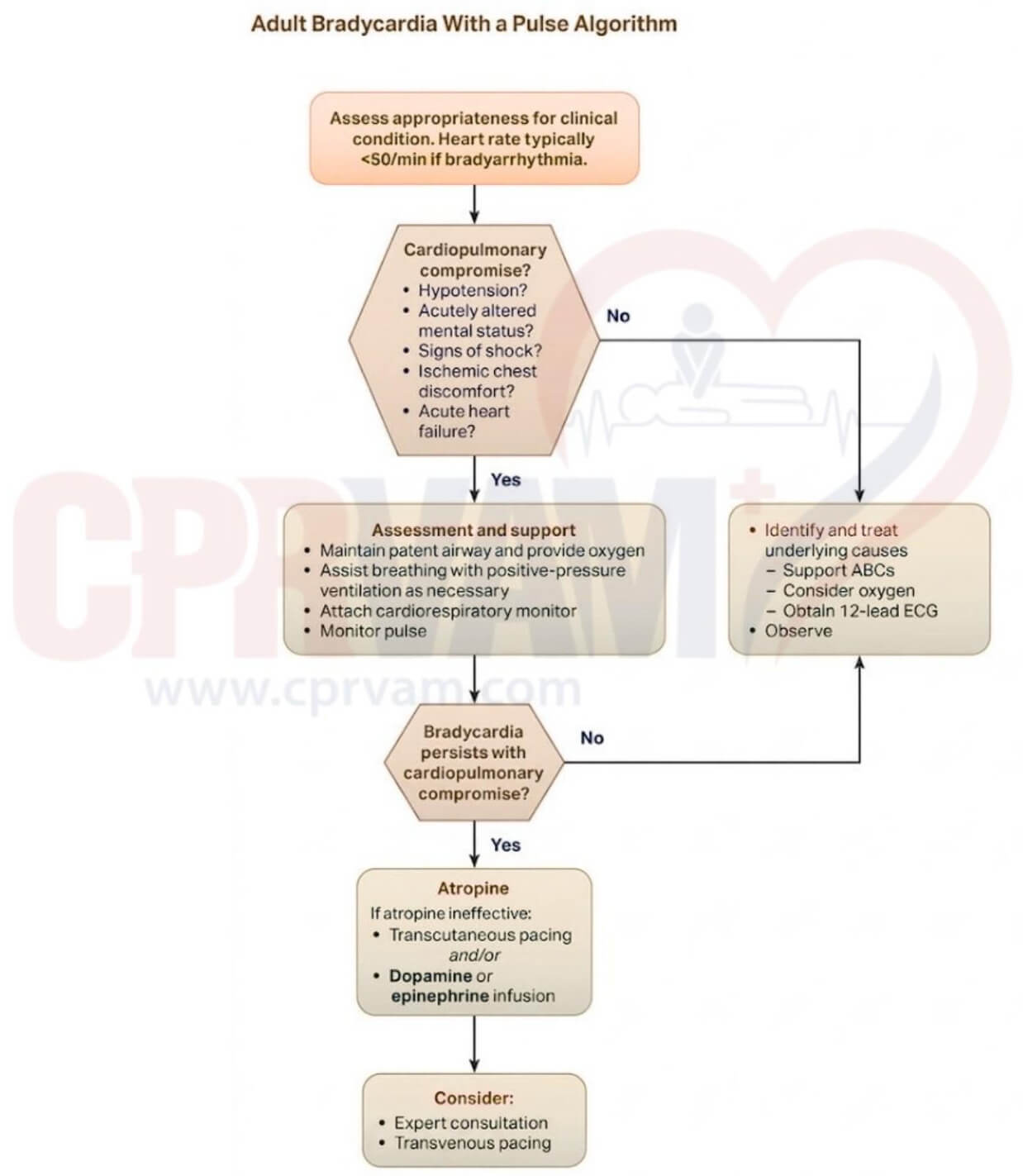
CPRvam Students
25,000+ students successfully trained annually
Step-by-step treatment algorithms
Adult bradycardia with pulse algorithm flowchart steps

It is a structured ACLS emergency approach for assessing and managing bradycardia with a pulse, guiding step-by-step evaluation, stabilization, symptom recognition, and timely treatment based on patient condition and response.

Here are the step-by-step Adult Bradycardia With a Pulse Algorithm:

The first step is recognizing clinically significant bradycardia and determining whether it is affecting circulation or organ function. Heart rate alone is not enough; symptoms and perfusion status are critical.

Clinical Priorities:

  • Recognize clinically significant bradycardia
  • Verify heart rate is typically below 50 beats per minute with symptoms
  • Assess impact on circulation and organ function

This step evaluates whether bradycardia is causing inadequate perfusion or organ dysfunction, helping determine if the patient is unstable and needs immediate intervention or can be monitored.

Clinical Indicators:

  • Hypotension
  • Acutely altered mental status
  • Signs of shock
  • Ischemic chest discomfort
  • Acute heart failure

The patient is considered unstable and requires immediate supportive intervention to maintain oxygenation and circulation.

Immediate Stabilization and Support:

  • Maintain patent airway and provide oxygen to ensure adequate oxygen delivery
  • Assist breathing with positive-pressure ventilation if respiratory effort is inadequate
  • Attach a cardiorespiratory monitor for continuous ECG and vital sign tracking
  • Monitor pulse continuously to assess perfusion and response to treatment

The patient is considered stable at this stage and does not require immediate emergency intervention. Focus shifts to evaluation and identifying reversible causes.

Monitoring and Diagnostic Evaluation:

  • Maintain airway patency and ensure adequate breathing and circulation
  • Administer oxygen when signs of hypoxia or respiratory distress are present
  • Perform a 12-lead ECG to evaluate rhythm abnormalities and possible ischemia
  • Investigate and manage reversible causes of bradycardia promptly

This phase focuses on maintaining oxygenation and circulation while preparing for definitive treatment. It helps prevent rapid deterioration during early evaluation.

Immediate Clinical Steps:

  • Ensure airway patency and protect the airway if the mental status is altered
  • Administer oxygen when hypoxia or respiratory distress is present
  • Assist ventilation if breathing becomes inadequate
  • Attach an ECG monitor and pulse oximeter for continuous tracking
  • Establish IV or IO access for emergency medications

This is the central decision point of the algorithm. It determines whether immediate intervention is required or if close observation is appropriate.

Unstable bradycardia is a serious condition where a slow heart rate causes inadequate blood flow to vital organs. It is identified by low blood pressure, altered mental status, signs of shock, chest pain from reduced heart perfusion, and features of acute heart failure or pulmonary congestion.

Management Priorities:

  • Secure the airway and provide oxygen support immediately
  • Assist ventilation if respiratory effort is insufficient
  • Maintain continuous ECG and hemodynamic monitoring
  • Ensure IV/IO access is ready for urgent drug delivery
  • Prepare for atropine administration and possible pacing
  • Reassess neurological status and perfusion frequently

Stable bradycardia is a condition where the heart rate is slow but blood circulation remains adequate, with preserved blood pressure, normal mental status, and no signs of chest pain, shock, or heart failure.

Response Approach:

  • Ongoing ECG observation for rhythm assessment
  • Obtain a 12-lead ECG for detailed evaluation
  • Provide oxygen only if clinically indicated
  • Perform frequent reassessments of vital signs
  • Focus on diagnostic workup and clinical observation

Atropine is the initial drug recommended by AHA guidelines for symptomatic bradycardia. It increases heart rate by blocking vagal (Parasympathetic) influence on the heart.

Medication Steps:

  • Administer Atropine 1 mg IV bolus
  • Repeat every 3-5 minutes if the response is inadequate
  • Maximum total dose: 3 mg
  • Monitor heart rate, blood pressure, and symptom improvement after each dose

If atropine fails or instability persists, additional therapies are required to maintain cardiac output and tissue perfusion.

Advanced Management Plan:

  • Transcutaneous pacing (external electrical cardiac stimulation)
  • Dopamine infusion at 5-20 mcg/kg/min, titrated to clinical response
  • Epinephrine infusion at 2-10 mcg/min for ongoing instability
  • Continuous ECG and hemodynamic monitoring during therapy

When bradycardia is persistent or severe, escalation to specialist care is necessary for long-term rhythm stabilization.

Clinical Direction:

  • Early involvement of cardiology or critical care teams
  • Consider transvenous pacing for definitive rhythm control
  • Transfer to the ICU for advanced monitoring and support
  • Adjust treatment plan based on ongoing patient response

Bradycardia can evolve quickly, so repeated evaluation is essential throughout care. Early detection of deterioration improves outcomes significantly.

Ongoing Evaluation Includes:

  • Continuous cardiac rhythm monitoring
  • Frequent checks of blood pressure and oxygen levels
  • Assessment of mental status and perfusion trends
  • Monitoring response to all interventions
  • Immediate escalation if the patient’s condition worsens

One of the key steps in the ACLS Bradycardia Algorithm is to identify, address, and treat the reversible causes using H’s and T’s mnemonic. Here are the symptoms and treatment for the H’s and T’s reversible causes of bradycardia:

Reversible Causes Symptoms Treatment
Hypoxia Shortness of breath, confusion, cyanosis, and organ dysfunction Managing the airway and adequate oxygenation
Hypovolemia Low blood pressure, rapid pulse, pale skin Includes rapid fluid resuscitation with crystalloids or blood products
Hydrogen ion (Acidosis) Respiratory acidosis from CO2 retention; metabolic acidosis from acid accumulation Correct the underlying cause, ventilation support, and sodium bicarbonate if metabolic
Hyperkalemia High potassium causes peaked T-waves, a widened QRS complex on ECG Calcium, insulin + glucose, bicarbonate, Kayexalate
Hypokalemia Low potassium causes flattened T-waves and prominent U-waves on ECG Controlled potassium infusion
Hypoglycemia Sweating, confusion, seizures IV glucose administration
Hypothermia Impaired metabolism, reduced drug response, arrhythmias Gradual rewarming, supportive care
Toxins (Drug Overdose, Poisoning) Include altered mental status, ECG changes, or abnormal heart rhythm Antidotes, supportive care, poison control consultation
Tamponade (Cardiac) Muffled heart sounds, jugular venous distension, hypotension Treatment by pericardiocentesis to drain fluid
Tension Pneumothorax Respiratory distress, absent breath sounds on the affected side, and distended neck veins Emergency needle decompression followed by chest tube insertion
Thrombosis (Coronary) Chest pain, dyspnea, hypoxia Anticoagulation, thrombolysis, supportive care
Thrombosis (Pulmonary Embolism) Severe chest pain, cough, fainting/syncope, leg swelling Anticoagulants, oxygen, IV fluids, thrombolysis/embolectomy
Trauma Absent pulses, low blood pressure, altered mental status, physical injury that may cause bleeding, organ damage, or airway obstruction Airway, breathing, circulation stabilization, hemorrhage control

The Adult Bradycardia With a Pulse Algorithm is a critical ACLS framework because it provides a structured, evidence-based approach to rapidly identify and manage symptomatic bradycardia before it progresses to cardiac arrest or severe hemodynamic collapse.

Key Importance Points:

  1. Rapid Recognition
    Enables early identification of clinically significant bradycardia by focusing on perfusion status and hemodynamic impact, not heart rate alone.

  2. Prevents Cardiac Arrest
    Promotes early detection of instability, allowing timely intervention before the patient deteriorates into cardiac arrest.

  3. Standardized Decision-Making
    Provides a clear, step-by-step clinical pathway for assessment, stabilization, and treatment in high-pressure emergency settings.

  4. Safe Treatment Selection
    Ensures correct use of ACLS interventions such as atropine, transcutaneous pacing, dopamine, and epinephrine based on patient response.

  5. Continuous Patient Safety Monitoring
    Emphasizes ongoing monitoring of ECG, blood pressure, mental status, oxygenation, and perfusion to detect early clinical changes.

  6. Improved Team Coordination
    Supports structured teamwork, clear roles, and timely escalation to advanced or specialist cardiac care when required.

  7. Better Survival Outcomes
    Increases the likelihood of recovery by ensuring rapid, guideline-based, and evidence-driven management of symptomatic bradycardia.

ACLS management of bradycardia requires careful clinical judgment, as treatment depends on the underlying cause, patient condition, and response to therapy rather than following the algorithm rigidly.

  1. Myocardial Infarction (MI)
    Bradycardia associated with acute MI requires cautious use of atropine, as it may be less effective and should not delay definitive reperfusion therapy and close monitoring.

  2. Do Not Resuscitate (DNR)
    Respect documented DNR orders. Avoid initiating resuscitative interventions such as pacing and instead focus on comfort-oriented and supportive care.

  3. Heart Transplant Patients
    Atropine is typically ineffective due to denervated cardiac tissue. Early consideration of pacing or alternative chronotropic support is recommended.

  4. High-Degree AV Block
    In cases of second-degree type II or third-degree AV block, especially with widened QRS complexes, atropine is often ineffective; early transcutaneous or transvenous pacing is preferred.

  5. Hypothermia
    Bradycardia may be physiologic in hypothermia. Prioritize active rewarming and avoid aggressive interventions like pacing until normothermia is achieved.

  6. Individualized Treatment
    ACLS algorithms provide a framework, but management should be tailored to the patient’s clinical condition and response to therapy, with continuous reassessment guiding decisions.

The ACLS Bradycardia Algorithm provides a clear, step-by-step approach for recognizing and managing symptomatic bradycardia using the most current AHA guidelines. It helps healthcare professionals assess the patient’s condition, identify signs of poor perfusion, and deliver timely, appropriate interventions, such as medications or pacing, to stabilize the heart rate.

That’s why anyone involved in advanced cardiac care needs to complete ACLS certification. The course offers in-depth knowledge and hands-on training in life-saving treatment algorithms, including the proper response to bradycardia and other cardiac emergencies. Learn critical skills with CPR VAM, an AHA-authorized training provider.

It is a stepwise emergency approach for patients with a slow heart rate, usually below 50 bpm, who show signs of poor perfusion. The focus is on identifying instability early and treating it before cardiac arrest develops.

Because a patient can have a slow heart rate but still be stable if perfusion is adequate. Treatment decisions depend on symptoms like blood pressure, mental status, and signs of shock rather than heart rate alone.

Patients are first assessed for cardiopulmonary compromise using signs such as hypotension, confusion, chest pain, shock, or heart failure. This determines whether they need immediate intervention or supportive monitoring.

The priority is stabilizing the airway, breathing, and circulation while initiating continuous monitoring. Oxygen support, ventilation assistance if needed, and IV/IO access are prepared without delay.

Because the patient’s condition can change quickly from stable to life-threatening. Ongoing monitoring of ECG, blood pressure, oxygen levels, and mental status ensures timely escalation of care when needed.

Advanced Cadiovascular Life Support