

Imagine being at home when a loved one suddenly collapses. You check their pulse, and it is gone, but the heart monitor shows normal electrical activity. This frightening scenario could be PEA Rhythms or Pulseless Electrical Activity (PEA), a critical type of cardiac arrest. Even though the heart seems to be working electrically, it is not pumping blood. Without immediate action, vital organs can fail, making rapid recognition and treatment essential for survival.
This blog breaks down PEA from start to finish. You will learn what it is, why it happens, and how to identify it on an ECG. It also explains life-saving ACLS treatments based on American Heart Association guidelines, including CPR, medications, and how to address the reversible causes known as the Hs and Ts. Understanding these steps can make the difference between life and death.
What Is Pulseless Electrical Activity (PEA)?
Pulseless Electrical Activity (PEA) happens when the heart’s electrical system appears to work, but the heart does not pump blood. You might see normal heart rhythms on a monitor, yet there is no pulse. Understanding PEA is crucial because it is life-threatening, and recognizing it quickly can help you act fast and save a life.
Types of PEA
There are two types of PEA:
True PEA
True PEA occurs when the heart’s electrical signals are present, but the heart muscle does not contract at all. You may see organized rhythms on the monitor, yet no blood moves through the body, creating an immediate emergency. Acting fast is critical to restore circulation.Pseudo-PEA
Pseudo-PEA happens when the heart muscles contract weakly, moving a small amount of blood, but not enough to generate a pulse. You might notice minimal blood flow, yet it is insufficient. Quick recognition and proper intervention can improve your patient’s chance of survival.
Characteristics of PEA Rhythms
Electrical Activity: The ECG shows organized electrical signals, but the heart muscle does not contract effectively.
Absence of Pulse: Despite the electrical activity, there is no detectable pulse or blood flow to the body’s organs.
Varied Rhythms: PEA can appear as a normal sinus rhythm or irregular patterns, but the heart still cannot pump blood.
No Corresponding Mechanical Contraction: The heart fails to contract mechanically, causing circulatory failure even though electrical signals are present.
Fact: PEA is a non-shockable rhythm, unlike Ventricular Fibrillation (VF) or Pulseless Ventricular Tachycardia (pVT). Defibrillation will not restore circulation; immediate CPR and cause-specific interventions are critical. (American Heart Association, 2025)
What Are the Main Causes of PEA & How Do the Hs and Ts Play a Role?
In most PEA cardiac arrests, the heart stops pumping due to reversible problems. The Hs and Ts framework explains these causes clearly, helping you recognize correctable conditions quickly and take the right life-saving actions.
PEA often results from reversible conditions, categorized by the “Hs and Ts” mnemonic:
1. Hs (Metabolic or Physiologic Causes)
Hypoxia: Low oxygen in the body can stop the heart from pumping, and you need to recognize it immediately.
Hypovolemia: Severe blood or fluid loss reduces circulation, and you must restore volume quickly.
Hydrogen Ion (Acidosis): A blood pH imbalance can impair heart function, so you should correct it promptly.
Hypo/Hyperkalemia: Too little or too much potassium disrupts heart rhythms, and you must manage it carefully.
Hypothermia: Dangerously low body temperature slows the heart, and you need to warm the patient safely.
Hypoglycemia: Low blood sugar can affect heart performance, so you should treat it without delay.
2. Ts (Mechanical or External Causes)
Tension Pneumothorax: A collapsed lung increases chest pressure, and you must relieve it quickly to restore circulation.
Tamponade (Cardiac): Fluid around the heart prevents proper pumping, and you need to drain it promptly.
Toxins: Drug overdoses, such as beta-blockers or calcium channel blockers, can stop the heart, and you should reverse or treat them.
Thrombosis: Blood clots in the lungs or heart block blood flow, and you must act fast to dissolve or bypass them.
Trauma: Severe injuries can cause circulatory failure, and you need to address the root cause immediately.
How Does PEA Differ From Other Cardiac Arrest Rhythms?
Cardiac arrest rhythms may look similar, but treatment decisions depend on key differences. Understanding how PEA compares with VF VT, and asystole helps you act fast, choose the right intervention, and follow ACLS guidelines correctly when a life is on the line.
Key Differences Between PEA and Other Cardiac Arrest Rhythms
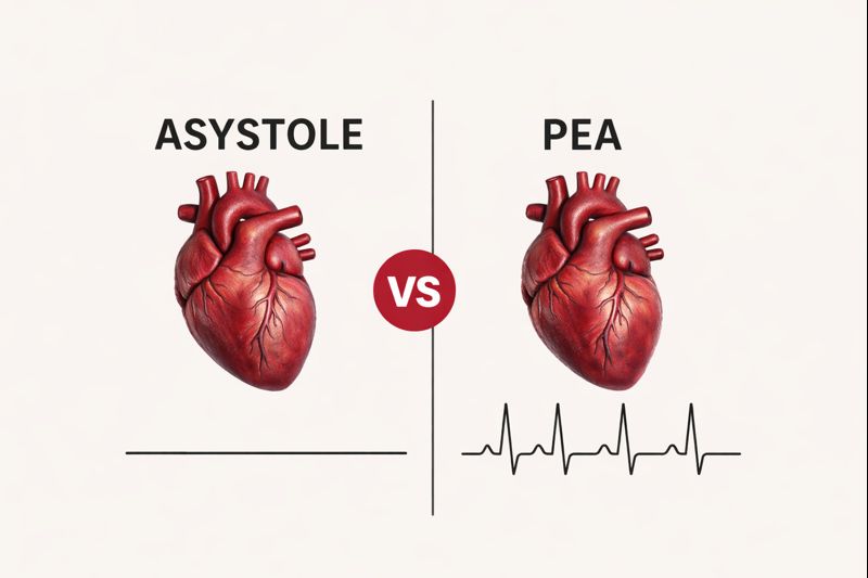
Features | Pulseless Electrical Activity (PEA) | Ventricular Fibrillation (VF) / Ventricular Tachycardia (VT) | Asystole |
Electrical Activity | An organized electrical rhythm is present | Chaotic or very rapid electrical activity | No electrical activity, flatline |
Pulse | No detectable pulse | No detectable pulse | No detectable pulse |
Shockable | No, defibrillation does not work | Yes, defibrillation is required | No, defibrillation is ineffective |
Primary Treatment | High-quality CPR, epinephrine, and treat reversible causes | Defibrillation, CPR, and medications | High-quality CPR and epinephrine |
Key Takeaway: Unlike VF or VT, PEA is not caused by faulty electrical activity. Instead, the heart cannot produce effective mechanical contraction.
What Clinical Signs Indicate Pulseless Electrical Activity?
Recognizing Pulseless Electrical Activity quickly can save a life. Even though the heart monitor shows electrical activity, the body receives no blood flow. Knowing the clinical signs helps you confirm PEA fast and begin the right ACLS actions without delay.
Here are the key signs that indicate PEA rhythms:
1. Check for Pulse
When you assess a patient in suspected PEA, you will find no palpable pulse despite organized electrical activity on the monitor. This mismatch between rhythm and circulation is a key sign that confirms cardiac arrest.
2. ECG Findings
On the ECG, you may see sinus rhythm, bradycardia, or another organized rhythm. Even though the tracing looks structured, you must remember that electrical activity alone does not mean the heart is pumping blood.
3. Clinical Signs of Cardiac Arrest
Patients in PEA are unresponsive and not breathing normally. You may also notice cyanosis or dilated pupils. These physical signs tell you the brain and organs are not getting oxygenated blood.
4. Immediate Threat
Because there is no effective circulation, PEA is a true medical emergency. Without rapid CPR, medications, and correction of reversible causes, organ damage and death can occur within minutes.
How Can You Treat PEA with Life-Saving ACLS Interventions?
Pulseless Electrical Activity is a life-threatening rhythm that requires rapid action. Understanding and following ACLS algorithms and interventions helps you provide high-quality CPR, administer medications, and address reversible causes, giving the patient the best chance for survival.
Step 1: High-Quality CPR
You must begin immediate chest compressions at 100-120 per minute, 5-6 cm deep. Allow full recoil after each push. High-quality CPR maintains blood flow to vital organs until circulation returns.
Essential Actions:
- Chest compressions 100-120/min
- Depth 5-6 cm for adults
- Full chest recoil
- Maintain a consistent rhythm
Step 2: Advanced Airway & Oxygenation
Secure the airway using an endotracheal tube or supraglottic device. Use capnography to monitor exhaled CO₂. You can confirm effective CPR and oxygen delivery when EtCO₂ exceeds 10 mmHg.
Critical Steps:
- Endotracheal or supraglottic airway
- Capnography monitoring
- EtCO₂ > 10 mmHg confirms perfusion
- Ensures proper oxygenation
Step 3: IV/IO Access and Medications
Establish IV or IO access to deliver life-saving drugs. Administer epinephrine every 3–5 minutes. Atropine, sodium bicarbonate, or thrombolytics may be needed depending on the underlying cause you identify.
Medication Checklist:
- IV/IO access established
- Epinephrine 1 mg every 3-5 min
- Atropine for bradycardia with hypotension
- Sodium bicarbonate for acidosis
- Thrombolytics for pulmonary embolism
Step 4: Identify and Correct Hs & Ts
Quickly find and treat reversible causes of PEA. You should oxygenate for hypoxia, drain a tamponade, relieve tension pneumothorax, and correct electrolyte imbalances to restore effective circulation.
Focus Areas:
- Treat hypoxia immediately
- Drain pericardial tamponade
- Relieve tension pneumothorax
- Correct electrolyte imbalances
- Focus on reversible causes
Step 5: Continuous Monitoring
Monitor the patient closely with repeated ECGs every 2 minutes. You watch for return of spontaneous circulation (ROSC) or changes in rhythm, adjusting interventions promptly to maximize chances of survival.
Monitoring Priorities:
- Repeat ECG every 2 minutes
- Watch for ROSC
- Observe rhythm changes
- Adjust treatment accordingly
Specialized Cardiac Intervention: In cases that do not respond to standard measures, advanced devices such as Extracorporeal Membrane Oxygenation (ECMO) or Intra-Aortic Balloon Pump (IABP) can be considered to maintain circulation.
How Can You Identify PEA Rhythm on An ECG?
You can identify Pulseless Electrical Activity (PEA) rhythm on an ECG by carefully observing the heart’s electrical patterns. Even if the rhythm appears organized, the heart may not produce an effective pulse, making recognition crucial for timely intervention.
Key Indicators of PEA Rhythm on ECG:
1. Organized Electrical Pattern
The ECG shows a rhythm that may look like a normal sinus rhythm or bradycardia, but you must remember that appearance does not guarantee effective heart pumping.
2. Absence of Mechanical Contraction
Despite visible electrical activity, there is no corresponding pulse, meaning the heart fails to circulate blood, and you need immediate action.
3. Resemblance to Other Rhythms
In severe cases, PEA may mimic idioventricular rhythm or even the transition to asystole, so you should carefully assess both the rhythm and the patient’s pulse.
Important Insight: An ECG may show electrical activity, but you cannot confirm blood flow without checking the patient’s pulse.
What Are the Most Common Mistakes When Managing PEA?
Understanding the most common mistakes when managing PEA helps you act effectively during cardiac arrest. Recognizing these errors ensures you deliver high-quality ACLS care, avoid delays, and address reversible causes promptly, improving patient outcomes.
Common Mistakes to Avoid:
1. Attempting Defibrillation on PEA
Never try defibrillation on PEA because it is a non-shockable rhythm. You should focus on high-quality CPR and correcting reversible causes for effective resuscitation.
2. Delaying High-Quality CPR
Immediate chest compressions are crucial. Any delay reduces oxygen delivery to vital organs, so you must start high-quality CPR without hesitation until circulation returns.
3. Ignoring Reversible Causes
Always identify and treat reversible Hs and Ts, like hypoxia, hypovolemia, or cardiac tamponade. Overlooking these factors can prevent successful resuscitation and decrease survival chances.
4. Skipping Continuous Monitoring
Continuously check the ECG and pulse every 2 minutes. Missing rhythm changes or the return of spontaneous circulation can delay critical interventions and worsen patient outcomes.
5. Neglecting Proper Airway Management and Oxygenation
Ensure the airway is secured and oxygen delivery is adequate. Poor ventilation can reduce oxygen to the heart and brain, undermining all other resuscitation efforts.
Strengthen Your Skills: Recognize, Respond, and Save Lives in PEA
Pulseless Electrical Activity is a critical cardiac arrest rhythm that requires rapid recognition and immediate action. Understanding the ECG patterns, delivering high-quality CPR, securing the airway, administering medications, and correcting reversible Hs and Ts are essential. Continuous monitoring ensures timely adjustments and improves survival chances. Mastering these interventions, you can confidently respond to PEA emergencies.
For healthcare providers aiming to boost their skills in handling cardiac emergencies like PEA, CPR VAM is here for you. Our AHA-approved courses in CPR, BLS, PALS, and ACLS provide hands-on guidance from expert instructors. Enroll today at a CPR VAM center near you and gain the confidence to act decisively in any cardiac arrest scenario.
FAQs About PEA
1. How Can You Recognize PEA?
You may notice the patient is unresponsive, has no detectable pulse, and shows abnormal breathing, even though the ECG displays an organized rhythm. Immediate CPR and emergency support are critical.
2. What Commonly Causes PEA?
PEA often results from severe blood loss (Hypovolemia), low oxygen (Hypoxia), acidosis, electrolyte imbalances, or mechanical problems such as cardiac Tamponade or Tension Pneumothorax.
3. How Is PEA Managed?
You should start high-quality CPR, give epinephrine every 3-5 minutes, provide oxygen, and quickly correct any reversible Hs and Ts.
4. Is PEA Shockable?
No. PEA is a non-shockable rhythm. Defibrillation is only effective for VF or VT.
5. What Does PEA Look Like on An ECG?
The ECG shows an organized rhythm that normally would generate a pulse, but there is no effective mechanical contraction, resulting in absent circulation.