



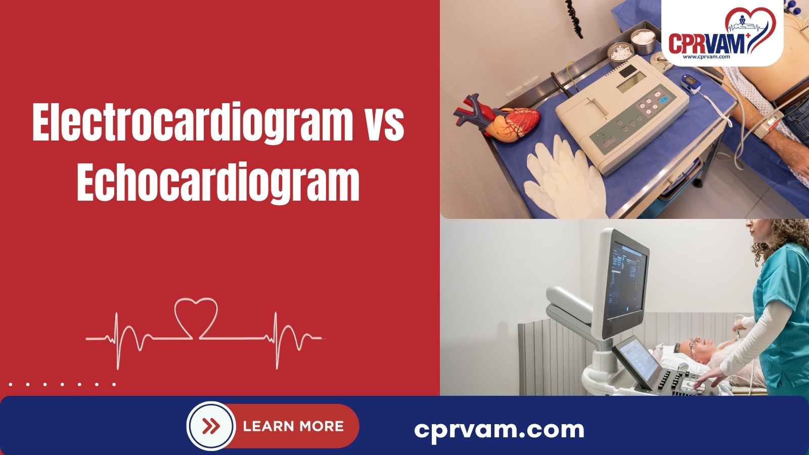
When your doctor orders a heart test, it is easy to confuse an Electrocardiogram (ECG or EKG) with an Echocardiogram (Echo). Electrocardiogram vs echocardiogram highlights a key difference: an ECG measures electrical activity, while an echocardiogram shows heart structure and function.
- An ECG Measures Electrical Activity: It records the electrical signals that control your heart’s rate and rhythm.
- An Echocardiogram Shows the Heart’s Structure and Function: It uses ultrasound to create moving images of the heart, including its chambers, valves, muscle, and blood flow.
Both tests provide essential information, but they answer different clinical questions. Depending on your symptoms, medical history, or initial findings, a healthcare professional may recommend one or both tests to build a complete picture of your cardiac health.
This blog explains the key differences between an ECG and an echocardiogram, including what each test measures, how it works, when it may be recommended, and what you can expect during the procedure.
ECG vs. Echocardiogram: Quick Comparison
The main difference between an ECG (electrocardiogram) and an echocardiogram (echo) is what they measure. An ECG records the heart’s electrical activity, while an echocardiogram uses ultrasound to show the heart’s structure and function.
Comparison Point | ECG (Electrocardiogram/EKG) | Echocardiogram (Echo) |
Primary Purpose | Evaluates the heart’s electrical activity | Evaluates the heart’s structure and function |
What It Measures | Heart rate, rhythm, and electrical signals | Heart chambers, valves, muscle movement, and blood flow |
Test Technology | Electrodes placed on the skin | Ultrasound waves and a handheld transducer |
Type of Result | Electrical tracing or waveform | Real-time images and measurements |
Common Clinical Uses | Identifies irregular heart rhythms and electrical abnormalities | Evaluates valve problems, structural abnormalities, and heart function |
How the Test Is Performed | Electrodes are attached to the chest and limbs | A transducer is moved across the chest |
Patient Experience | Generally painless and quick | Generally painless; mild pressure may be applied |
Invasiveness | Noninvasive | Noninvasive for a standard echocardiogram |
Best For Evaluating | The heart’s electrical system | The heart’s structure, movement, and pumping function |
Typical Test Duration | About 5-10 minutes | About 30-60 minutes, depending on the type |
Type of Result | Electrical patterns and measurements | Images and measurements of heart structure and function |
What Is an Electrocardiogram (ECG/EKG)?
An electrocardiogram (ECG or EKG) is a quick, noninvasive test that records the heart’s electrical activity. It helps healthcare professionals check the heart rate, rhythm, and electrical signals, and identify signs of certain heart problems.
What Does an ECG Measure?
An ECG measures the electrical signals that control each heartbeat. It can provide information about:
- Heart Rate: How fast the heart is beating
- Heart Rhythm: Whether the heartbeat is regular or irregular
- Electrical Activity: How electrical signals move through the heart
- Abnormalities: Patterns that may indicate an electrical or heart-related problem
- Possible Heart Damage: Changes that may suggest previous or ongoing heart damage
How Does an ECG Work?
During an ECG, small adhesive electrodes are placed on the chest and sometimes on the arms and legs. These electrodes detect electrical signals and send them to an ECG machine, which displays the heart’s activity as waveforms on a tracing.
An ECG does not send electricity into your body. Instead, it records the electrical signals naturally produced by your heart. These signals help healthcare professionals assess your heart’s rhythm and identify potential electrical abnormalities that may require further evaluation.
What Happens During an ECG?
An ECG is quick and painless. You will lie still while a healthcare professional places small electrodes on your skin. The machine records your heart’s electrical activity for a short time, helping check your heart rhythm and identify possible problems.
You may feel slight discomfort when the electrodes are removed, especially if they pull on body hair. However, the test does not send electricity into your body or cause an electric shock. It is generally safe and simple.
When Is an ECG Recommended?
A healthcare professional may recommend an ECG when they need to evaluate symptoms or monitor a known heart condition. Common reasons include:
- Chest pain or discomfort
- Heart palpitations
- Irregular heartbeat
- Dizziness or fainting
- Shortness of breath
- Possible heart attack
- Abnormal heart rhythms
- Monitoring certain heart conditions
- Checking pacemaker function or treatment response
An ECG may also be included as part of a routine evaluation when a healthcare professional believes it is appropriate.
What Are the Different Types of ECG Tests?
Different ECG-based tests can monitor the heart under different conditions:
- Resting 12-Lead ECG: Records the heart’s electrical activity while you are resting.
- Exercise or Stress ECG: Records heart activity while you exercise, usually on a treadmill or stationary bike.
- Holter Monitoring: Uses a portable device to continuously record heart activity, often for 24 hours or longer.
- Event Monitoring: Records heart rhythm over a longer period and may be activated when symptoms occur.
- Specialized ECG Monitoring: Other forms of monitoring may be used depending on the symptoms or heart condition being evaluated.
What Is an Echocardiogram?
An echocardiogram (echo) is a noninvasive heart test that uses ultrasound waves to create moving images of the heart. Unlike an ECG, which records the heart’s electrical activity, an echocardiogram helps healthcare professionals see how the heart is structured and how well it is working.
What Does an Echocardiogram Measure?
An echocardiogram provides images and measurements that can help evaluate:
- Heart chambers and their size
- Heart muscle and movement
- Heart valves and how they open and close
- Pumping function and how well the heart moves blood
- Wall motion and movement of the heart muscle
- Blood flow through the heart
- Structural abnormalities that may affect heart function
How Does an Echocardiogram Work?
An echocardiogram uses a handheld device called a transducer to send high-frequency sound waves into the body. These waves bounce back as they interact with the heart and nearby tissues, allowing the machine to collect information about the heart.
The echocardiography system processes these returning sound waves into real-time images of the heart. These images help healthcare professionals examine the heart’s structure, movement, and function while it beats, making it easier to identify potential problems.
What Happens During an Echocardiogram?
For a standard transthoracic echocardiogram (TTE), you lie on an examination table while a healthcare professional applies gel to your chest. They gently move the transducer across different areas of your chest to capture images of your heart.
A standard echocardiogram is generally painless and noninvasive. You may feel slight pressure as the healthcare professional moves the transducer across your chest to capture clear images of your heart’s structure and movement during the test.
When Is an Echocardiogram Recommended?
A healthcare professional may recommend an echocardiogram when they need more information about the heart’s structure or function. Common reasons include evaluating:
- Heart valve problems
- Heart failure or reduced pumping function
- Heart muscle abnormalities
- Enlarged heart chambers
- Structural heart conditions
- Heart murmurs
- Certain congenital heart conditions
- Abnormal heart movement or function
An echocardiogram may also be used to monitor a known heart condition and assess how the heart responds to treatment.
What Are the Types of Echocardiograms?
Different types of echocardiograms are used depending on what needs to be evaluated:
- Transthoracic Echocardiogram (TTE): The most common type, performed by moving a transducer over the chest.
- Transesophageal Echocardiogram (TEE): Uses a specialized transducer placed in the esophagus to obtain detailed images of the heart.
- Stress Echocardiogram: Combines echocardiography with exercise or medication that increases the heart’s workload.
- 3D Echocardiogram: Produces three-dimensional images that can provide additional detail about the heart’s structure and function.
- Specialized Echocardiographic Studies: Other techniques may be used when a healthcare professional needs more specific information.
ECG vs. Echocardiogram: Key Differences Explained
An ECG and echocardiogram both evaluate the heart, but they provide different information. An ECG focuses on electrical activity, while an echocardiogram uses ultrasound to examine the heart’s structure and function.
Here are the key differences between an ECG and an Echocardiogram:
1. What Each Test Evaluates
An ECG records the heart’s electrical activity, helping healthcare professionals evaluate heart rate, rhythm, and electrical signals. It can identify patterns that may suggest an irregular heartbeat or other electrical abnormalities affecting normal heart function.
An echocardiogram creates moving images of the heart, helping healthcare professionals evaluate its chambers, valves, muscle movement, pumping function, and blood flow. It can also identify structural abnormalities that may affect how the heart works.
2. Technology Used for Each Test
An ECG uses small electrodes attached to the skin to detect electrical signals produced by the heart. The electrodes send these signals to a machine, which records them as electrical waveforms for interpretation.
An echocardiogram uses a handheld transducer and ultrasound waves to create images of the heart. The transducer sends sound waves into the body and receives returning signals that are processed into real-time images.
3. Results and Findings
An ECG produces a waveform tracing that shows the heart’s electrical activity. A healthcare professional performs an EKG interpretation to assess heart rate, rhythm, and electrical conduction, including the PQRST wave pattern and other waveform changes that may require further evaluation.
An echocardiogram produces moving images and measurements of the heart. A healthcare professional can use these results to assess heart structure, valve function, muscle movement, pumping ability, and blood flow.
4. Procedure and Patient Experience
During an ECG, small electrodes are placed on the skin, usually on the chest and sometimes the arms and legs. You typically remain still while the machine records your heart’s electrical activity.
During a standard echocardiogram, gel is applied to the chest before a transducer is moved over different areas. The test captures images while the heart beats and is generally painless and noninvasive.
Note: The simplest way to remember the difference is that an ECG shows how the heart’s electrical system is working, while an echocardiogram shows the heart’s structure, movement, and pumping function.
When an ECG and Echocardiogram Are Used Together
An ECG and echocardiogram provide different types of heart information. An ECG records electrical activity, including heart rate and rhythm, while an echocardiogram uses ultrasound to assess the heart’s structure, valves, movement, and pumping function. Using both tests can help healthcare professionals evaluate different aspects of heart health.
A doctor may recommend both tests when symptoms, medical history, or initial findings require further evaluation. For example, an ECG may identify an abnormal rhythm, while an echocardiogram can help assess whether a structural or functional heart problem may be contributing to the findings. The choice depends on individual clinical needs.
ECG and Echocardiogram for Common Heart Conditions
The role of an ECG and echocardiogram can vary depending on the heart condition being evaluated. An ECG primarily assesses the heart’s electrical activity, while an echocardiogram provides information about heart structure and function. Healthcare professionals may use one or both tests based on symptoms, medical history, and clinical findings
1. Abnormal Heart Rhythms
An ECG is commonly used to evaluate abnormal heart rhythms because it records the heart’s electrical activity. If a rhythm problem occurs intermittently, longer-term ECG-based monitoring may help capture changes that may not appear during a short test.
An echocardiogram may also be recommended to evaluate the heart’s structure and function. It can help identify structural or functional abnormalities that may be associated with certain abnormal heart rhythms.
2. Reduced Heart Function
An ECG can provide information about heart rhythm and electrical conduction and may help identify changes that require further evaluation. However, it does not directly show how well the heart muscle pumps blood.
An echocardiogram can assess heart structure and pumping function, including how the heart chambers and muscle are working. This makes it an important test when evaluating reduced heart function or possible heart failure.
3. Heart Valve Conditions
An echocardiogram is particularly useful for evaluating heart valve conditions. It can show the structure and movement of the valves and help assess problems such as valve narrowing or leakage.
An ECG may also be performed when valve disease affects heart rhythm or places additional strain on the heart. The two tests can therefore provide different but complementary information.
4. Chest Pain and Suspected Heart Attack
An ECG may be used to evaluate chest pain and identify electrical changes that can occur during a heart attack or other cardiac conditions. It is often one of the initial tests used when a heart-related cause of chest pain is suspected.
Depending on the symptoms and clinical findings, healthcare professionals may also order blood tests, imaging, or other cardiac tests. The appropriate evaluation depends on the individual situation and suspected cause.
Are ECGs and Echocardiograms Safe?
A standard ECG and echocardiogram are generally considered safe, noninvasive tests. An ECG records the heart’s electrical activity through electrodes placed on the skin, while a standard transthoracic echocardiogram uses ultrasound waves to create images of the heart. Neither test uses ionizing radiation.
A standard ECG does not send electricity into the body; it only records the heart’s natural electrical signals. A standard echocardiogram is also generally painless, although you may feel some pressure from the transducer. Specialized echocardiograms, such as a transesophageal echocardiogram (TEE), involve additional considerations because the probe is passed through the mouth into the esophagus.
How Long Do ECG and Echocardiogram Tests Take?
A standard ECG (electrocardiogram/EKG) typically takes about 5-10 minutes in total. The actual recording takes only a few seconds, while the remaining time is generally used to position and attach the electrodes to the chest and limbs and prepare for the test.
A standard echocardiogram usually takes about 30-60 minutes. During the test, a healthcare professional applies gel to the chest and moves a handheld transducer over different areas to capture images of the heart’s chambers, valves, movement, and function. The exact time may vary by test type.
ECG vs Echocardiogram: Which Test Is Better?
Neither test is always better than the other. An ECG and echocardiogram provide different information about the heart, so the right test depends on what a healthcare professional needs to evaluate.
- ECG → Evaluates the heart’s electrical activity and rhythm
- Echocardiogram → Evaluates the heart’s structure, movement, and function
- Both → Provide complementary information when clinically appropriate
The appropriate test depends on factors such as symptoms, medical history, physical examination findings, and the specific heart concern being evaluated. A healthcare professional can determine which test, or combination of tests, is most appropriate.
Choose the Right Heart Test for Your Needs
ECG and echocardiogram tests provide different but valuable information about the heart. An ECG helps assess electrical activity and rhythm, while an echocardiogram provides images of the heart’s structure and function. Depending on your symptoms, medical history, and examination findings, a healthcare professional may recommend one or both. Understanding their roles can help you know what to expect and discuss appropriate testing with your healthcare professional.
For those looking to build practical skills for responding to cardiac emergencies, CPR VAM Training Center provides BLS, ACLS, PALS, and Heartsaver CPR courses to help participants develop essential emergency response skills and confidence.
FAQs: Electrocardiogram vs Echocardiogram
1. Is an ECG the Same as an Echocardiogram?
No. An ECG and an echocardiogram are different heart tests. An ECG records the heart’s electrical activity, including its rate and rhythm. An echocardiogram uses ultrasound to create images of the heart’s structure and function.
2. Can an Echocardiogram Detect Heart Problems?
Yes. An echocardiogram can help evaluate several aspects of heart health. It can assess the heart chambers, valves, muscle movement, pumping function, and blood flow. It may also help identify certain structural abnormalities and heart conditions.
3. Can You Have an ECG and Echocardiogram on the Same Day?
Yes. Both tests can be performed on the same day when clinically appropriate. The timing may depend on the healthcare setting, symptoms, and reason for testing. Your healthcare professional can explain how the tests will be scheduled.
4. Is an Echocardiogram More Detailed Than an ECG?
Neither test is simply more detailed because they examine different aspects of the heart. An echocardiogram provides images of heart structure and function, while an ECG provides information about electrical activity and rhythm. The test that provides more useful information depends on the clinical question.
5. Does an Echocardiogram Replace an ECG?
No. An echocardiogram does not replace an ECG. The two tests evaluate different aspects of heart health and may provide complementary information. A healthcare professional may recommend one or both based on your symptoms and clinical needs.