


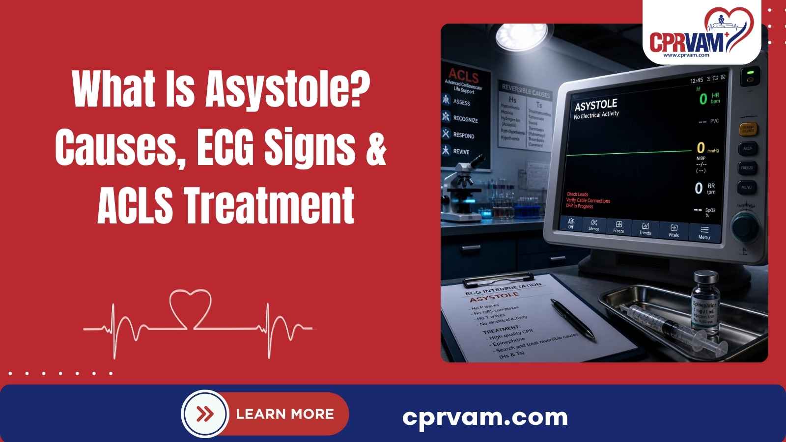
A sudden flatline on a cardiac monitor can signal a life-threatening emergency. But what is asystole, and why does it require immediate action? Asystole is a cardiac arrest rhythm with no detectable electrical activity. As a non-shockable rhythm, it requires immediate CPR, epinephrine, airway and ventilation support, and treatment of reversible causes.
A flatline on a cardiac monitor can signal asystole, a life-threatening cardiac arrest rhythm with no detectable electrical activity. Rapid recognition and treatment are critical. Trained rescuers should begin high-quality CPR, follow the appropriate ACLS algorithm, and identify reversible causes to improve the chance of restoring circulation.
This blog explains what asystole is, what causes it, how it appears on an ECG, how it differs from PEA, and what to do during an asystolic cardiac arrest.
What Happens to the Heart During Asystole?
During asystole, the heart has no detectable electrical activity, so it cannot produce effective contractions or pump blood. As a result, blood flow to the brain and other vital organs stops. Asystole is therefore a form of cardiac arrest that requires immediate recognition and resuscitation to restore circulation.
A suspected flatline should always be confirmed before identifying it as asystole. Check the patient, ECG leads, electrode connections, and monitoring equipment to rule out technical problems. Loose electrodes or disconnected leads can create a false flatline. Once asystole is confirmed, begin appropriate resuscitation and address potentially reversible causes promptly.
How Does the Heart Work?
The heart is a muscular pump that uses electrical signals to coordinate each heartbeat. These signals help the heart contract in the right sequence, allowing blood to move efficiently to the lungs and the rest of the body.
- Electrical Impulse: The sinoatrial (SA) node initiates the heartbeat.
- Atrial Contraction: The atria contract and move blood into the ventricles.
- Ventricular Contraction: The ventricles contract and pump blood to the lungs and body.
- Relaxation: The heart relaxes and fills with blood before the next heartbeat.
This coordinated process keeps blood circulating throughout the body. In asystole, detectable electrical activity is absent, so the heart cannot produce effective contractions or maintain circulation.
What Causes Asystole?
Asystole occurs when the heart loses detectable electrical activity, usually because of a severe underlying condition. During cardiac arrest, ACLS providers look for reversible causes of asystole, commonly grouped into the Hs and Ts. Identifying and treating these causes quickly is an important part of effective cardiac arrest management.
1. The Hs: Reversible Causes
- Hypovolemia: Severe blood or fluid loss can reduce circulation and contribute to cardiac arrest.
- Hypoxia: A lack of oxygen can impair heart function and lead to cardiac arrest.
- Hydrogen ion (acidosis): Severe acidosis can interfere with normal cardiac function.
- Hypo-/Hyperkalemia: Abnormal potassium levels can disrupt the heart’s electrical activity.
- Hypothermia: Severe hypothermia can slow body processes and contribute to cardiac arrest.
2. The Ts: Reversible Causes
- Tension Pneumothorax: Trapped air in the chest can restrict circulation and reduce blood return to the heart.
- Cardiac Tamponade: Fluid or blood around the heart can prevent the chambers from filling properly.
- Toxins: Certain drugs, medications, or poisons can interfere with cardiovascular function.
- Thrombosis: A coronary or pulmonary blood clot can severely restrict blood flow.
3. Additional Factors/Other Causes
Asystole can also occur after severe heart disease, myocardial infarction, drowning, trauma, electrocution, sepsis, or prolonged hypoxia. In some cardiac arrests, prolonged ventricular fibrillation (VF) or pulseless ventricular tachycardia (pVT) can eventually deteriorate into asystole.
Because some causes are reversible, ACLS providers should continue evaluating the Hs and Ts during resuscitation. Treating the underlying cause, while maintaining high-quality CPR and following the appropriate ACLS protocol, can be critical to restoring circulation.
What Are the Signs of Asystole/Cardiac Arrest?
Asystole occurs during cardiac arrest and is typically accompanied by signs that indicate the heart is no longer pumping blood effectively. Recognizing these signs quickly is critical because every second matters during a cardiac emergency.
Common signs of asystole and cardiac arrest include:
- Unresponsiveness: The person does not respond to voice or touch.
- Absent or Abnormal Breathing: Normal breathing is absent, or the person may have occasional gasping.
- No Detectable Pulse: There is no pulse when assessed by a trained responder.
- Loss of circulation: The heart is not effectively pumping blood to the brain and other vital organs.
- Flat or Nearly Flat ECG: The monitor may show no detectable electrical activity.
If cardiac arrest is suspected, activate the emergency response system and begin high-quality CPR immediately. Trained healthcare professionals should follow the appropriate resuscitation protocol and assess the cardiac rhythm to guide further treatment.
How Is Asystole Identified on an ECG?
Asystole appears on an ECG as a flat or nearly flat line with no detectable cardiac electrical activity. But what does a flat line mean? While it may indicate asystole, a flatline alone does not confirm the rhythm. Healthcare professionals should assess the patient and verify the monitoring equipment before confirming asystole.
What Should You Check?
When a suspected flatline appears on the monitor:
- Assess the Patient: Check responsiveness, breathing, and signs of circulation.
- Check the Electrodes: Make sure ECG leads and electrode connections are secure.
- Verify the Rhythm: Confirm the tracing in another lead when appropriate.
- Check the Equipment: Ensure the monitor is connected and functioning correctly.
- Confirm the rhythm: Distinguish asystole from other cardiac arrest rhythms, particularly PEA.
This verification helps prevent technical problems, such as disconnected leads or loose electrodes, from being mistaken for true asystole. Accurate rhythm identification is essential because asystole is a non-shockable rhythm and requires a different treatment approach from shockable rhythms such as ventricular fibrillation.
Quick Reference: Asystole vs. Other Cardiac Arrest Rhythms During ECG
Rhythm | ECG Finding | Pulse | Shockable? |
Asystole | No detectable electrical activity | Absent | No |
VF | Chaotic, disorganized ventricular activity | Absent | Yes |
pVT | Rapid ventricular rhythm | Absent | Yes |
PEA | Organized electrical activity | Absent | No |
Accurate rhythm identification is essential because cardiac arrest treatment differs between shockable and non-shockable rhythms.
Asystole vs. PEA: What's the Difference?
Asystole and pulseless electrical activity (PEA) are both non-shockable cardiac arrest rhythms, but they differ in what the ECG shows. Knowing the difference helps healthcare professionals identify the rhythm and follow the appropriate ACLS treatment pathway.
Feature | Asystole | PEA |
Electrical activity | No detectable cardiac electrical activity | Organized electrical activity is present |
Pulse | Absent | Absent |
Effective contraction | No effective cardiac contraction | No effective pulse despite electrical activity |
Defibrillation | Not indicated | Not indicated |
CPR | Required | Required |
Reversible causes | Identify and treat | Identify and treat |
The Key Difference
- Asystole: No detectable cardiac electrical activity.
- PEA: Electrical activity is present, but there is no palpable pulse or effective circulation.
Because both rhythms are non-shockable, treatment focuses on high-quality CPR, epinephrine according to ACLS protocols, and identifying and treating reversible causes.
What Immediate ACLS Actions Are Required for Asystole?
Asystole is a non-shockable cardiac arrest rhythm that requires immediate resuscitation. ACLS management focuses on high-quality CPR, timely epinephrine, appropriate airway and ventilation support, and identification of reversible causes. Because asystole is not treated with defibrillation, accurate rhythm recognition is essential for choosing the appropriate treatment approach.
1. Start High-Quality CPR
Begin high-quality CPR immediately when cardiac arrest is recognized. Effective chest compressions help maintain blood flow to the heart and brain while the resuscitation team provides additional treatment. Keep interruptions to a minimum and allow complete chest recoil. Continue CPR while other ACLS interventions are performed and the rhythm is reassessed.
2. Administer Epinephrine as Early as Possible
For non-shockable rhythms such as asystole, administer epinephrine as soon as possible after IV or IO access is established. Continue high-quality CPR during medication administration and repeat epinephrine every 3 to 5 minutes according to ACLS protocols. At the same time, identify and treat reversible causes to support effective resuscitation.
3. Manage the Airway and Ventilation
Provide appropriate oxygenation and ventilation throughout resuscitation. Bag-mask ventilation may be used initially, while an advanced airway can be considered when appropriate and performed by trained professionals. Avoid excessive ventilation and coordinate airway management with chest compressions. The approach should support oxygen delivery without unnecessarily interrupting high-quality CPR.
4. Identify and Treat Reversible Causes
Continue evaluating the Hs and Ts throughout resuscitation because some causes of asystole may be reversible. The team should consider conditions such as hypoxia, hypovolemia, electrolyte abnormalities, tension pneumothorax, cardiac tamponade, thrombosis, and toxins. Promptly identifying and treating the underlying cause can be critical to restoring circulation.
5. Do Not Defibrillate Asystole
Confirmed asystole is a non-shockable rhythm, so defibrillation is not indicated. Immediate ACLS management focuses on high-quality CPR, epinephrine, airway and ventilation support, and identifying and treating reversible causes. Defibrillation is reserved for shockable rhythms such as ventricular fibrillation (VF) and pulseless ventricular tachycardia (pVT).
6. Reassess the Rhythm
Reassess the cardiac rhythm at appropriate intervals during CPR, typically during rhythm checks associated with 2-minute CPR cycles. If the rhythm becomes shockable, follow the appropriate shockable-rhythm algorithm. If return of spontaneous circulation (ROSC) occurs, transition to post-cardiac-arrest care and continue monitoring and treatment as indicated.
What Happens After ROSC?
Return of spontaneous circulation (ROSC) means that the heart has regained sustained circulation after cardiac arrest. However, ROSC is not the end of resuscitation. The patient still needs close monitoring and post-cardiac-arrest care to support organ function, identify the cause of the arrest, and reduce the risk of further complications.
What Does Post-ROSC Care Include?
After ROSC, healthcare professionals may focus on:
- Oxygenation and Ventilation: Support adequate oxygen levels and appropriate ventilation while avoiding both hypoxia and excessive oxygenation.
- Blood Pressure and Circulation: Maintain adequate blood pressure and support circulation to protect vital organs.
- Cardiac Evaluation: Obtain a 12-lead ECG and assess for the underlying cause of cardiac arrest.
- Neurological Monitoring: Monitor neurological status and assess for complications following cardiac arrest.
- Temperature Control: Use an appropriate, protocolized temperature-control strategy when indicated, particularly in patients who remain unresponsive after ROSC.
- Treating the Underlying Cause: Identify and address reversible causes or other conditions that contributed to the cardiac arrest.
- Ongoing Critical Care: Continue close monitoring and provide appropriate intensive care based on the patient’s condition.
The specific approach depends on the patient’s clinical status, cause of cardiac arrest, neurological condition, and response to treatment. Current AHA guidelines emphasize that post-cardiac-arrest care is an essential part of the resuscitation process, not simply the next step after ROSC.
What Are the Survival Chances for Asystole?
Survival after asystolic cardiac arrest is generally poor compared with shockable cardiac arrest rhythms. However, outcomes vary based on the cause of arrest, time to treatment, CPR quality, the patient’s condition, and whether a reversible cause can be corrected.
Factors That Affect Outcomes
- Time to intervention: Early recognition and immediate CPR help maintain blood flow to the brain and heart.
- Underlying cause: Identifying and treating a reversible cause may improve the possibility of restoring circulation.
- Quality of CPR and ACLS: High-quality CPR, appropriate medication administration, airway management, rhythm reassessment, and treatment of reversible causes are important during resuscitation.
- Response to treatment: Whether ROSC occurs and how the patient responds to post-cardiac-arrest care also affect the overall outcome.
The cardiac rhythm is only one factor; overall outcomes depend on the patient’s condition and response to treatment.
Take Action to Improve Survival Chances
Asystole is a medical emergency that requires immediate recognition and resuscitation. Trained responders should begin high-quality CPR, activate the emergency response system, follow the appropriate ACLS algorithm, administer epinephrine when indicated, support ventilation, and identify reversible causes. Understanding what asystole is, what a flatline may indicate, and why confirmed asystole is not treated with defibrillation can help responders provide an appropriate cardiac arrest response.
Staying current with ACLS training can help healthcare professionals respond confidently during cardiac emergencies. CPR VAM’s ACLS courses provide hands-on training designed to strengthen essential resuscitation skills.
Train today and be prepared to respond when every second matters.
FAQs About Asystole
1. What Does Flat Line Mean on an ECG?
A flat or nearly flat ECG line may indicate asystole, but it does not confirm the rhythm by itself. Check the patient, ECG leads, electrode connections, and monitoring equipment to rule out technical problems. Accurate rhythm verification is important before confirming asystole and determining the appropriate treatment.
2. Can You Defibrillate or Shock Asystole?
No. Confirmed asystole is a non-shockable rhythm, so defibrillation is not indicated. Treatment focuses on high-quality CPR, epinephrine according to ACLS protocols, airway and ventilation support, and identifying and treating reversible causes, including the Hs and Ts, during resuscitation.
3. Is Asystole the Same as a Flatline?
Asystole typically appears as a flat or nearly flat line on an ECG because there is no detectable cardiac electrical activity. However, a flatline should be verified before it is identified as true asystole. Loose electrodes, disconnected leads, or equipment problems can produce a false flatline.
4. Can Asystole Be Reversed?
Asystole may sometimes be reversed when a treatable underlying cause is identified and addressed quickly. Immediate CPR, appropriate ACLS treatment, and correction of reversible causes may help restore return of spontaneous circulation (ROSC). However, asystolic cardiac arrest generally has a poor prognosis.
5. Can Young People Experience Asystole?
Yes. Asystole can occur in people of any age when a severe condition leads to cardiac arrest. Potential causes include trauma, drowning, severe hypoxia, poisoning, or serious cardiac disease. Rapid recognition, CPR, and appropriate emergency treatment are critical regardless of age.